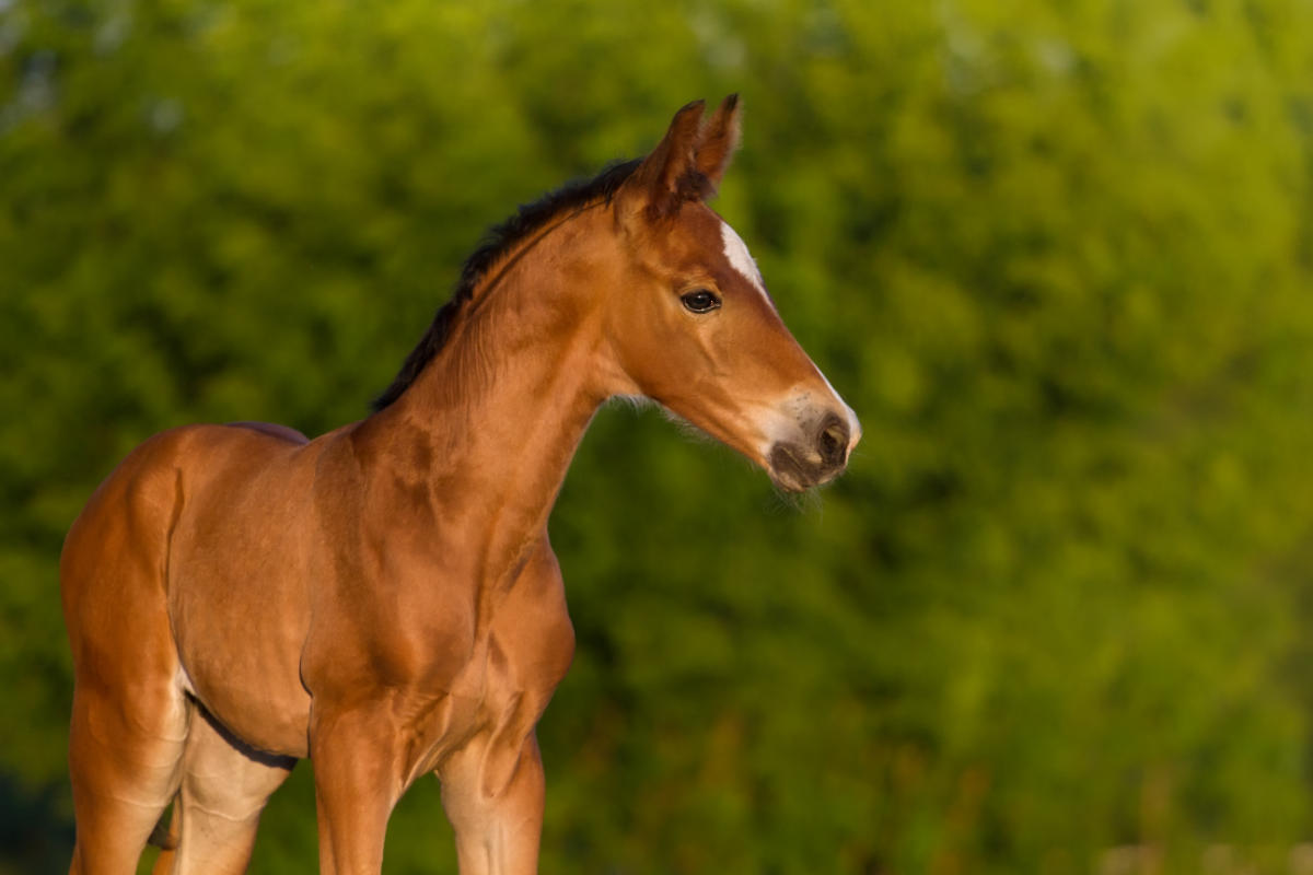
Juvenile osteochondral disease (J OCD) is a complex group of disorders affecting the development of cartilage and subchondral bone in young horses. These conditions, often grouped under the umbrella term osteochondrosis, are of major concern to breeders and veterinarians because of their significant impact on the health and future performance of equines. AOJ can manifest as lameness, joint deformity and, in the most severe cases, permanent disability. Understanding the underlying mechanisms, risk factors and prevention approaches is essential to ensure the well-being and optimal development of young horses.
What causes this disease?
Juvenile osteochondral disease (JOCD) can seriously compromise a horse’s sporting career from the very first months of life. These disorders, which include abnormalities in the ossification process, particularly affect foals. JCOA encompasses various types of lesions, including :
- Osteochondrosis dissecans (OCD): characterised by irregularities or fragmentation of the bone surfaces and the formation of subchondral bone cysts.
- Bone tears: resulting from the traction of a ligament.
These anomalies are often caused by biomechanical stresses on the foal’s immature skeleton. They lead to disturbances in blood flow to the ossification site. The economic consequences for the equine industry are significant, as they reduce the commercial value of the foal and its future sporting performance.
The clinical signs of AOCJ vary according to the severity of the lesions, the joint affected, the location of the lesion within the joint, and the horse’s use (leisure versus competition). Lesions in the tibio-tarsal joints may be subclinical, while those in the stifle are often symptomatic.
Environmental and individual factors play a crucial role in the development of AOCJ. Inappropriate feeding of the pregnant mare and the growing foal, as well as excessive biomechanical stress, can favour these conditions. In addition, rapid growth and physical conformation influence the prevalence of lesions. Genetic factors are also a determining factor, with some breeds being more predisposed to these disorders than others.
What are the symptoms of osteochondrosis?
As with many diseases, the symptoms of osteochondrosis can vary from one horse to another, depending on the location of the lesions and the type of work the animal does. For some horses, osteochondrosis may cause no discomfort at all, or only at certain times, while for others the symptoms may be more pronounced and visible.
The most common symptoms include:
- Swelling of the affected joint: This is one of the most common signs, resulting from inflammation of the joint. This swelling, due to an increase in the amount of synovial fluid, may not be painful for the horse but indicates the presence of a bone fragment (or chip).
- Lameness: The severity of lameness varies from one horse to another and is often most noticeable during work. It may be caused by pain linked to distension of the joint or a bone cyst.
- Joint pain: The affected joint may be sensitive to manipulation, especially during flexion. The pain may be caused by synovial distension or a fragment wedged between the bones of the joint.
Osteochondrosis is often asymptomatic in young, untrained horses, but may manifest itself with symptoms such as:
- Swelling of the joint
- Pain on passive mobilisation of the affected joint
- A positive flexion test
- Lameness of varying degrees
How is it diagnosed?
Osteochondrosis may be diagnosed as a preventive measure or if there is clinical suspicion. The initial clinical examination helps to guide the diagnosis by palpation of the joints and flexion tests of the limbs to detect reduced range of movement or pain.
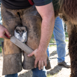
Veterinarians often supplement the clinical examination with a dynamic examination to refine the diagnosis. They use diagnostic local anaesthetics to localise pain precisely. They usually confirm lesions using imaging, in particular radiographic examinations . Veterinarians may also use ultrasound images to obtain additional information.
The ESOAP study demonstrated the evolution of AOCJ in foals between 6 and 18 months of age, indicating that the lesions evolve significantly during this period. Radiographic screening around the age of one year is recommended to get a precise idea of the foal’s osteoarticular status.
Your vet will begin with a full locomotor examination, looking at the limbs for swelling, sensitive areas or areas of increased heat. He will then observe the horse in movement and carry out flexion tests to check for any lameness. If necessary, additional examinations such as radiography or ultrasound will be carried out to locate and assess the severity of the lesions.
What treatments are available?
Treatment of osteochondrosis depends on the severity of the lesions, the joint affected and the horse’s use. There are two main types of treatment available: conservative treatment and surgical treatment.
Conservative treatment involves rest and the administration of anti-inflammatories. It is appropriate when the lesions are minor or if the horse is not intended for intense sporting activity. The aim of this treatment is to adapt the horse’s activity to avoid aggravating the lesions and to manage the symptoms with anti-inflammatories and possibly infiltrations.
Arthroscopic surgery is often recommended for more severe cases. This technique involves making small incisions to insert a camera and surgical instruments to remove bone fragments or debride cysts. Arthroscopy, performed under general anaesthetic, also enables the overall health of the joint to be assessed. Surgery is generally of short duration. It has a low complication rate. What’s more, it offers a favourable prognosis for young horses destined for a sporting career.
In the case of subchondral cysts, several surgical techniques can be used to fill the cyst and prevent it from expanding. However, the high cost of these operations encourages some owners to opt for conservative treatment.
What are the natural alternatives?
For young growing horses, it is crucial to support their ossification period to ensure good bone quality. Feed supplements such as Ekyflex Osteo can be beneficial. For horses with fragments or who have had fragments, the use of chondroprotectors can help protect the joints.Harpagophytum-based supplements such as Harpagyl are also useful for preventing stiffness.
Thanks to its anti-inflammatory active ingredients,harpagophytum is effective in treating the pain associated with osteoarthritis. Extracts need to be taken over several months to see the beneficial effects.
White willow bark is also used for its analgesic and anti-inflammatory properties. Containing salicylin, which is transformed into salicylic acid in the body, it acts by inhibiting the enzymes responsible for inflammation.
Boswellia, turmeric and blackcurrant are other plants with anti-inflammatory and analgesic effects. They are traditionally used to treat osteoarticular pain. Incorporating them into horses’ diets can help manage the symptoms of osteochondrosis naturally.
How can osteochondrosis be prevented?
Preventing osteochondrosis in horses involves monitoring a number of factors, including diet, physical activity and early detection.
The diet of the broodmare and foal is crucial. The diet should be balanced in terms of energy and protein, with particular attention paid to protein quality and mineral and trace element intake. An imbalance in calcium and phosphorus or a copper deficiency can increase the risk of osteochondrosis. The use of mineral and vitamin supplements (CMV) may be necessary to make up for dietary deficiencies.
Regular moderate physical activity is important for the healthy development of the foal’s skeleton. We recommend avoiding sudden transitions between phases of inactivity and intense activity. Permanent grazing, rather than stabling, can help to maintain regular physical activity without excess.
Early screening enables lesions to be detected and treated quickly. This avoids having a negative impact on the horse’s future sporting career. X-ray screening is recommended between 18 and 24 months of age, especially for horses with a genetic predisposition. Early screening increases the chances of effective treatment and a good sporting prognosis.
Avoiding breeding individuals with a history of osteochondrosis and finding out about the osteoarticular status of the parents can reduce the risk. Moderate, regular exercise during growth, combined with a balanced diet with no abrupt dietary transitions, are key factors in preventing this pathology.