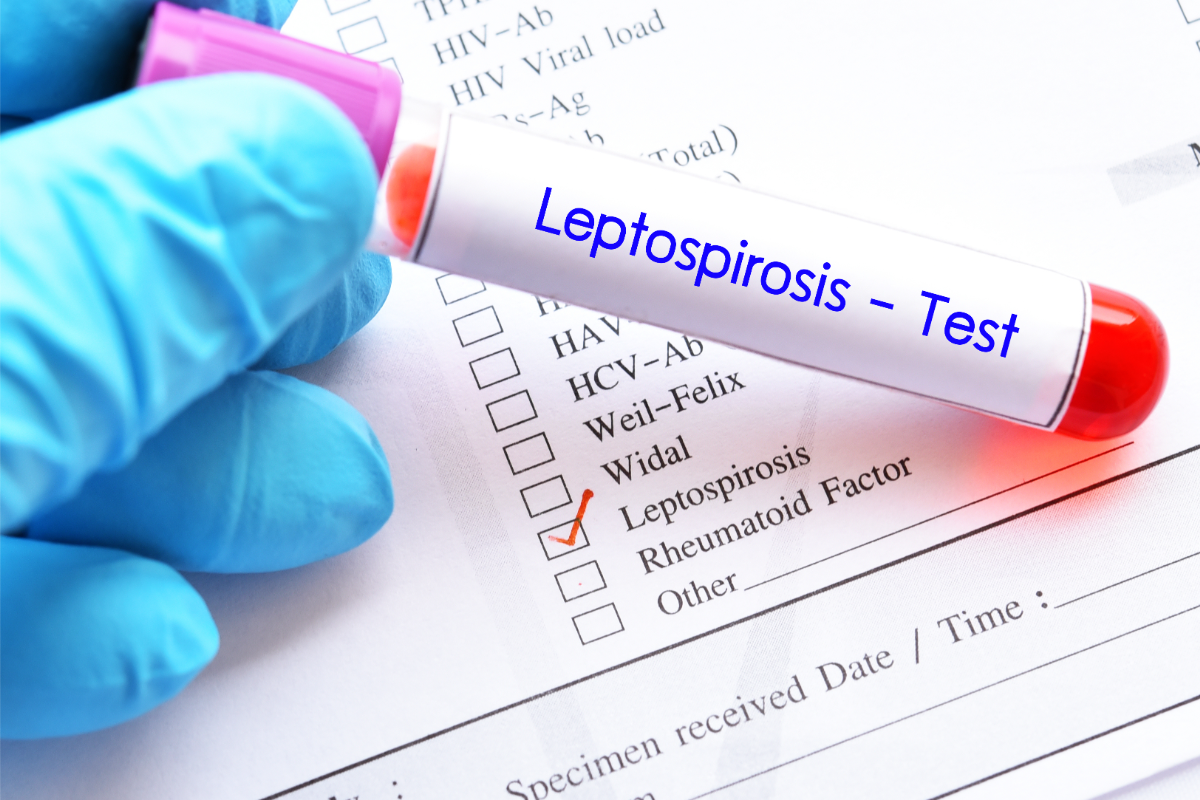
Leptospirosis is a bacterial infectious disease that mainly affects animals, but can also affect humans. Caused by bacteria of the genus Leptospira, this zoonosis is often underestimated despite its potentially serious implications for public health. It is transmitted mainly through exposure to water or soil contaminated by the urine of infected animals, particularly rodents.
What bacterium is responsible?
Leptospirosis is an infectious disease of varying severity, caused by bacteria of the genus Leptospira, of the order Spirochetes. The pathogenic bacteria of the Leptospira genus include several species, including Leptospira interrogans, for which there are more than 250 varieties known as serovars. The most common in mainland France are L. icterohaemorrhagiae, L. australis, L. sejroe and L. grippotyphosa.
These are anthropozoonoses, diseases common to humans and animals (mammals). The main reservoirs are wild rodents (healthy carriers), followed by dogs and livestock (pigs, horses, cattle). The Leptospira genus also includes saprophytic, non-pathogenic species, such as L. biflexa, which live in soil and freshwater and do not infest animals. In the course of evolution, certain species have adapted to the kidney tubules of mammals. They cause leptospirosis in humans and domestic animals.
Leptospira interrogans is the pathogen responsible for leptospirosis. In 2017, the Leptospira genus comprised 22 species, including 10 pathogens, and more than 300 serovars divided into 24 serogroups. The most important serogroups are icterohaemorragiae, canicola, pomona, australis, grippotyphosa, hyos and sejroe. This diversity makes it difficult to design effective vaccines against all leptospiroses.
Leptospira bacteria are between 6 and 20 micrometres long and 0.1 micrometres in diameter. It is aerobic and grows slowly at 27-30°C on special media. It appears as a spiral filament, flexible and mobile, with hooked ends and a terminal endoflagellum consisting of a pair of periplasmatic flagella. This structure gives it mobility and speed. This facilitates its diffusion into tissues and its ability to evade immune defence mechanisms such as phagocytosis.
Since the beginning of the 21st century, a new classification based on the genome has tended to complement the traditional classification based on antigens.
What does the disease look like in animals?
Leptospirosis is an infectious disease caused by pathogenic bacteria of the genus Leptospira. It is widespread throughout the world, with a particularly high incidence in tropical areas. In Europe, the situation varies from country to country.
All mammals can be infected by leptospires. Susceptible species, such as livestock, pets and hobby animals, can develop disease, the dog being the most susceptible species. Species with little or no susceptibility, mainly rodents, can excrete the bacteria via their urine. Some susceptible species, such as beavers and foxes, may harbour the bacteria, although their susceptibility is not well known.
Transmission occurs mainly through contact of mucous membranes or injured skin with fresh water, soil or an environment contaminated by the urine of infected animals. Leptospires can survive for several weeks in freshwater. Transmission can also occur via contaminated biological fluids, mainly the urine of infected animals.
Clinical signs vary from species to species. In dogs, liver and kidney damage can cause death within a few days without treatment. Horses, cattle and pigs may show reproductive problems. Rodents are generally asymptomatic, with the exception of beavers, hamsters and guinea pigs, but are kidney carriers.
In dogs, leptospirosis is mainly due to Leptospira canicola and L. interrogans. Infected dogs rapidly develop fever and vomiting. Treatment is based on antibiotic therapy. A veterinary vaccine is available, although its effectiveness may be limited against certain leptospiroses.
In cattle and sheep, leptospirosis can cause economic losses for farmers by affecting reproduction and milk production. In horses, ocular complications can occur.
How is it transmitted?
Leptospirosis is transmitted by contact with mucous membranes or injured skin. It occurs mainly through exposure to fresh water, soil or an environment contaminated by the urine of infected animals, where leptospires can survive for several weeks. Contaminated biological fluids, particularly urine, are also transmission vectors.
Occupations at risk include work in contact with freshwater or damp ground soiled by rodent urine (rats, coypu, muskrats and mice). Sewage workers, water treatment plant staff, river bank maintenance workers, fish farmers, fisheries wardens and workers in aquatic environments are particularly at risk. Breeders, vets, slaughterhouse workers, renderers and animal handlers who come into contact with domestic rodents may also be at risk.
The bacterium generally enters the skin . It seems to be facilitated by a break in the skin or skin softened by prolonged exposure to water. Contamination can also occur through the mucous membranes by splashing soiled water into the mouth, nose or eyes. Inhalation of aerosols is less common. Leisure activities such as swimming, fishing, kayaking and rafting also increase the risk of transmission.
After penetrating the skin or mucous membranes, leptospires pass into the bloodstream and migrate to all tissues. The incubation period and severity of infection depend on the quantity of leptospires inoculated.
Leptospires, present in water following the droppings of contaminated animals, enter the body through wounds, skin erosions, mucous membranes or by inhalation. Humans are occasional hosts of pathogenic leptospires, which cycle between wild and domestic animals.
How does leptospirosis manifest itself in humans?
The incubation period for leptospirosis varies from 1 to 3 weeks, usually between 7 and 14 days. The disease presents a number of clinical forms. These can range from flu-like symptoms to multivisceral disease with haemorrhagic syndrome, making diagnosis difficult. The fatality rate is between 5% and 20% in the absence of treatment or if treatment is delayed. Leptospirosis is typically biphasic, but can sometimes be monophasic and fulminant. Diagnosis is more likely in summer, when people are exposed to freshwater or riverbanks for work or leisure.
The two phases of leptospirosis
Leptospirosis begins with a septicaemic phase, in which the leptospires multiply in the blood and damage the small blood vessels through vasculitis. This causes severe fever and widespread pain. Next, the leptospires attach themselves to various organs (liver, kidney, brain, heart), making diagnosis difficult. This phase begins abruptly, with headaches, intense myalgias, chills, fever, cough and sometimes haemoptysis. The eyes turn red, usually around the 3rd or 4th day. The septicaemic phase lasts from 4 to 9 days.
After the septicaemic phase, an immune phase occurs between days 6 and 12, marked by the appearance of antibodies in the serum. Symptoms reappear, with renewed fever and signs of meningitis. Ocular disorders such as iridocyclitis, optic neuritis and peripheral neuropathy may also occur. Pulmonary complications can be severe in cases of pulmonary haemorrhage. This phase generally lasts from 4 to 30 days.
Renal involvement leads to excretion of leptospires in the urine, but this occurs too late to aid diagnosis. In humans, there are no healthy carriers, and they are not a reservoir of transmission.
If infected during pregnancy, leptospirosis can lead to miscarriage, even during convalescence. The disease in humans can be severe if left untreated, with a case-fatality rate of 5% to 20%. Severe forms can include renal failure, neurological disorders (convulsions, coma) and potentially fatal haemorrhage.
It is vital to consult a doctor if you develop a fever within two weeks of swimming or playing in freshwater, and to report any such activity.
Classic form: Weil’s disease
Jaundice-haemorrhagic leptospirosis or Weil’s disease is a classic clinical form of leptospirosis. It combines jaundice and renal damage (hepatonephritis) with haemorrhagic disorders. Leptospira interrogans, mainly serovar icterohaemorrhagiae, causes the disease, although other serovars are possible.
Once considered the most serious form of leptospirosis, specialists now see Weil’s disease as a more common and less serious form in developed countries.
Clinical manifestations vary in intensity. In the majority of cases, they are not very serious, but can develop unpredictably into severe disorders with a mortality rate of over 10%.
The onset is sudden, with a severe infectious syndrome (high fever, chills, asthenia) and pain (headache, muscle pain). Jaundice appears after 2 to 8 days, reaching its peak in 2 days, producing a “flamboyant jaundice”. This phase lasts around 10 days, followed by a lull and possible relapse.
Renal damage, characterised by albuminuria and reduced urine production, may progress to acute renal failure requiring haemodialysis. Bleeding disorders include petechiae, epistaxis and bleeding gums, with frequent thrombocytopenia. Serious haemorrhage may occur in the respiratory, digestive and urogenital tracts.
Rare complications such as meningitis and multiple organ damage (cardiovascular, pulmonary, septic shock) may require specific resuscitation. Weil syndrome can lead to fever, jaundice, renal failure and haemorrhagic tendencies. The lungs and heart may also be severely affected.
In the absence of jaundice, recovery is complete. In the presence of jaundice, the fatality rate is 5-10%, rising to 40% in severe cases, especially in patients over 60. The risk of death increases with complications such as renal failure, respiratory failure and internal haemorrhage.
Flu-like form
The flu-like form of leptospirosis, which accounts for around 80% of cases, is more common than the classical form. The onset of the disease is sudden, with a severe infectious state: fever over 39°C, chills, headaches, accompanied by myalgias and diffuse pain predominantly in the lower limbs, particularly incapacitating calf pain, aggravated by pressure.
Clinical examination may reveal several distinctive signs, including conjunctival haemorrhage, herpes labialis and sometimes painful hepatomegaly. More rarely, an exanthema may appear on the trunk. This initial phase of the disease lasts from 3 to 7 days, during which time the fever gradually returns to normal.
If left untreated, a relapse may occur. The initial symptoms reappear, generally less intense, but accompanied by meningeal signs and neurological disorders. These signs include a stiff neck, severe headaches, and sometimes more serious symptoms such as convulsions. Ocular complications may also appear later, in particular uveitis of immunological origin, caused by autoantibodies.
The initial phase of influenza-like illness is marked by high fever, severe muscle pain, headache and chills. These symptoms can mimic those of influenza. This makes clinical diagnosis difficult without a history of exposure to leptospirosis risk factors.
The flu-like form of leptospirosis is characterised by an initial severe but often non-specific presentation. It is then followed by a possible relapse with neurological and ocular complications. This anicteric form of leptospirosis is the most common. It accounts for the majority of cases, and requires particular vigilance for early diagnosis and treatment.
Other forms
What is the appropriate treatment?
The standard treatment for leptospirosis is a penicillin antibiotic (penicillin G or ampicillin) or a cyclin such as doxycycline. This is a probabilistic antibiotic treatment that needs to be started early, lasting 7 to 10 days. This early antibiotic treatment has all but eliminated serious chronic forms of the disease, particularly auto-immune ocular complications.
In the case of visceral and metabolic complications, resuscitation methods may be necessary, such as dialysis for persistent renal failure. A Jarisch-Herxheimer reaction, due to lysis of the spirochetes, is common after the start of treatment.
The patient recovers within 5 to 6 weeks if the disease is moderate. However, bacteria may still be present in the urine several weeks after symptoms have disappeared. Severe forms of leptospirosis have a mortality rate of over 10% worldwide, but in countries with modern medical infrastructures, mortality is close to zero.
For severe forms, doctors administer corticosteroids, although their effectiveness is open to debate. Antibiotic treatment is administered early for maximum effectiveness. Isolation is not necessary, but precautions must be taken to eliminate urine.
Treatment of severe forms requires hospitalisation with medical resuscitation and antibiotics administered as soon as possible. Third-generation cephalosporins (ceftriaxone and cefotaxime) and azithromycin are the first-line treatments. Cyclins may be offered in cases of allergy. Leptospires are usually sensitive to β-lactams, macrolides and cyclins.
A vaccine (SPIROLEPT®) is available for the prophylaxis of leptospirosis due to the Icterohaemorrhagiae serogroup in adults at high risk.
What are the means of prevention?
Actions at reservoir level include:
- Rodent control: deratting, elimination of food and shelter sources, management of rubbish in closed containers, design of premises.
- Environmental management: drainage of wet meadows, elimination of stagnant water.
- Health surveillance: declaration and management of abortions on farms.
- Isolation and treatment of sick animals: if animals are kept, curative treatment.
- Animal vaccination: adapted to the species concerned.
To limit transmission, it is necessary to :
- Limit contact with fresh water in areas frequented by rodents.
- Avoid all direct contact with wild animals, whether alive or dead.
- Organise work sites: identify rodents and damp areas if working in damp or infested environments.
- Safe transport: of waste and cadavers in sealed, labelled containers.
- Cleaning and disinfection: of contaminated sites and reusable service equipment with an authorised bactericide.
- Hygiene facilities: separate lockers for street clothes and work clothes, drinking water, soap, single-use wiping materials, and a first-aid kit agreed with the occupational physician.
- Information on recruitment and regularly updated
- In the laboratory, follow good practice in accordance with current regulations.
For individual prevention , we rely on :
- Personal protective equipment: resistant, waterproof gloves, boots or waders, waterproof overalls, protective goggles depending on the activity.
- Compliance with hygiene instructions
Laboratories in France prepare the SPIROLEPT® vaccine from inactivated L. interrogans serovar Icterohaemorrhagiae bacteria. This vaccine targets adults at high risk. The vaccination schedule includes two injections 15 days apart, a booster 4 to 6 months later, and then every 2 years if exposure persists.
In veterinary medicine, dogs are generally vaccinated against four serogroups: L. Canicola, L. Icterohaemorrhagiae, L. Australis and L. Grippotyphosa.
Some epidemiological data…
Leptospirosis is not a compulsory animal disease (Regulation 2016/429). However, it is a notifiable human disease. The authorities recognise leptospirosis as an occupational disease for which compensation is payable under Table 5 of the agricultural scheme and Table 19 of the general scheme. The authorities classify all Leptospira interrogans serovars in group 2 (article R.4421-3 of the French Labour Code, Order of 16 November 2021).
Leptospirosis is a worldwide disease, with a particularly high incidence in tropical regions. In mainland France, it affects around 600 people every year (0.4 to 1/100,000 inhabitants). The incidence is 50 to 100 times higher in tropical regions. It is estimated that more than a million severe cases occur each year worldwide, with a mortality rate in excess of 10%. The disease is highly seasonal, with peaks during the rainy season in tropical regions and in summer/autumn in temperate countries.
Certain professions (farmers, stockbreeders, sewage workers, refuse collectors) and water-based leisure activities (swimming, canoeing, kayaking, fishing, hunting, canyoning) present increased risks.
Epidemiology varies according to ecosystem and living conditions. In France, the number of cases has risen from 300 to around 600 per year since 2014. The southern regions and Franche-Comté are the most affected. In French overseas departments, the incidence is 10 to 80 times higher than in mainland France.
There are many reasons for this emergence: global warming, increased rodent populations and high-risk activities. The systematic reporting of cases of leptospirosis since August 2023 aims to better assess the disease, identify populations at risk, and implement appropriate control measures.


