Sarcoids are the most common skin tumour in horses, representing a significant challenge for owners and vets. These benign but persistent lesions can appear anywhere on the animal’s body, affecting quality of life and, in some cases, performance. This article explores in depth the different forms of sarcoid, diagnostic methods, available treatment options and prevention strategies. By providing comprehensive and up-to-date information, we aim to equip horse owners and animal health professionals with the knowledge they need to effectively manage this condition.
What is sarcoid?
Equine sarcoid, defined in 1936, is a locally invasive but non-metastatic mesenchymal and fibroblastic skin tumour. It accounts for 1 to 2% of equine consultations, affecting around 1 in 10 horses during its lifetime. Sarcoids account for 35-90% of skin neoplasia diagnosed in horses, donkeys, mules and zebras.
Sarcoids can occur in horses of any age, with a higher prevalence between the ages of 2 and 9. Sex has no significant influence, although geldings are slightly more affected than stallions. The breeds most affected are Appaloosa, Quarter Horse and Paint Horse, while Lipizzaners and Trotters are less affected.
There are six types of sarcoid:
- Occult: benign, alopecic area of skin.
- Verrucous: dry, horny skin mass.
- Nodular: firm subcutaneous nodules (subtypes A and B).
- Fibroblastic: fleshy, ulcerated, very complicated.
- Mixed: combination of several types.
- Malignant: aggressive, invasive, may infiltrate lymph nodes.
Sarcoids often recur after treatment, with a recurrence rate of 20% to 80%. They frequently develop on sites of old wounds, possibly linked to a genetic factor and the presence of bovine papillomavirus. Regular monitoring is essential for early diagnosis and effective management.
Microscopically, sarcoids show a proliferation of palisade-like fibroblasts at the dermal-epidermal junction, with a hyperplastic epidermis. According to the WHO, sarcoid is a mesenchymal tumour specific to horses, differing from fibrosarcoma in its biphasic component. This feature is pathognomonic and is investigated by the histopathologist.
Why does my horse have sarcoids?
There are several main causes of sarcoidosis in horses:
- Skin lesions: Sarcoids often develop on scars, poorly healed wounds or following insect bites.
- Viruses: Bovine papillomavirus (BPV) is suspected of playing a role in the development of sarcoids. Studies have shown that this virus may be involved in their development.
- Genetic predisposition: Certain breeds such as Quarter Horses, Appaloosas and Arabian horses show a genetic predisposition to sarcoids. A small wound on a genetically susceptible horse, bitten by an insect carrying the virus, can lead to the appearance of sarcoids.
BPV can be transmitted by direct or indirect contact (brushes, blankets) and via insects. BPV mainly infects dermal fibroblasts. Oncoproteins such as E5, E6 and E7 play a crucial role in tumorigenesis.
Sarcoids rarely regress spontaneously and can progress rapidly following trauma. The viral genome has been found in sarcoids, but no intact virions.
Research shows variations in the BPV genome and possible transmission by insects. Genetic studies indicate moderate heritability and chromosomal regions associated with sarcoid.
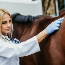
How do vets distinguish sarcoids from other tumours?
Even with a favourable clinical examination, sarcoids are difficult to diagnose. Although histological diagnosis is the gold standard, new approaches have been developed. A diagnostic protocol improves the sensitivity and specificity of clinical diagnosis, based on a scoring system similar to the “sepsis score”. A high score probably indicates sarcoid, while a low score suggests another tumour. A biopsy is recommended in the case of a medium score to optimise case selection, reducing expenditure and the risk of the lesion recurring after excision.
The role of bovine papillomavirus (BPV) in the genesis of sarcoids has been proven, and screening tools using PCR and ISH are effective. However, some studies have shown that BPV can be detected in samples of healthy skin. A recent study revealed that the latency phase between infection and clinical expression was short.
A new method of fine needle aspiration sampling has been developed, increasing the detection of BPV in epithelium-intact sarcoids. It is crucial to rule out other hypotheses when faced with a skin mass, in particular schwannoma, which is often histologically confused with sarcoid. Differential diagnosis is based on the appearance, location and evolution of the lesions, with confirmation by histological analysis.
What are the treatment options?
Treatment must take into account the aesthetic or functional damage caused by sarcoid. Despite their local aggressiveness, treatment remains difficult due to their invasive nature and tendency to recur. Depending on the number and type of tumour, its location, the cosmetic or functional damage, and the financial resources available, several types of treatment may be considered. Some sarcoid tumours may regress spontaneously without treatment.
Invasive treatments
Surgical excision is frequently used to control the growth of sarcoids. It is a rapid and cosmetically effective method. Techniques include simple excision,laser excision,electrosurgery and cryosurgery.
- Simple excision: Performed by ligature or under general anaesthetic, this method is often followed by frequent recurrences. Ligation uses a rubber band to cut off the blood supply, but is ineffective for extensive sarcoids.
- Laser excision: Precise and minimising bleeding, this technique has a non-recurrence rate of 83%. It works by vaporising the intracellular water in neoplastic tissue.
- Electrosurgery: This technique, which requires a general anaesthetic, combines excision and coagulation using alternating current. It has a success rate of 86.8%.
- Cryosurgery: Uses liquid nitrogen to destroy neoplastic tissue by forming intracellular ice. Success rates vary between 70% and 80%.
Surgical methods must be chosen according to a number of factors, including the location and size of the tumours, the veterinary surgeon’s skills and the financial resources available. Laser therapy is an effective alternative with a low recurrence rate. Cryosurgery is also effective, although less so recently in favour of laser surgery. Early intervention is crucial to reduce recurrence and improve long-term results.
Non-invasive treatments
Topical treatments for sarcoid are mainly in the form of ointments, offering ease of use and good compliance. However, it is essential to distinguish between scientifically validated products and others. Here’s an overview of the options:
- Imiquimod ointment: This ointment, with anti-tumour and anti-viral properties, induces the activity of cytokines (interferon-α, tumour necrosis factor-α). Studies show cure rates of up to 84.4%, but it can cause side effects such as swelling, erosions and ulcerations.
- Aciclovir ointment: Used against herpesviruses, its efficacy against sarcoids remains limited. An initial study showed complete regression in 68% of cases, but subsequent research found no significant difference compared with a placebo.
- Electrochemotherapy: This method combines intratumoral administration of platinum-based anti-cancer drugs with electrical pulses. Success rates without recurrence are as high as 97.9% after four years.
- Immunotherapy: The BCG vaccine, historically used, has shown inconsistent results. Feline interleukin-2, used for feline fibrosarcoma, has shown promising results against sarcoid, although further confirmation is required.
- Photodynamic therapy: Non-invasive, this method uses photosensitive substances under targeted lighting to treat small, non-invasive sarcoids, with promising results.
- Radiotherapy: Effective but costly, this includes strontium plesiotherapy, brachytherapy and teletherapy, with high success rates but cost and accessibility constraints.
- Tigilanol tiglate: Used for canine mastocytomas, it is also effective against sarcoids, with complete regression after a single intratumoral injection.
The choice of sarcoid treatment should be based on proven efficacy, potential side effects and the specific circumstances of each case.
Phyto-aromatherapy
Sanguinaria canadensis is combined with zinc chloride in ointments such as Xxterra® or Redbalm®. Bloodroot contains alkaloids with antibacterial, antifungal, anti-inflammatory and antiplatelet properties. It suppresses angiogenesis and induces cell death by breaking viral DNA strands. A study by Pettersson et al (2020) showed a cure rate of 75% after daily application of the ointment for 6 days, then every 4 days until complete remission, or for a maximum of 45 weeks. The best results were observed on small sarcoids.
These ointments, although intended for veterinary use, are available over the counter on the Internet. Legislation in France has restricted their availability.
The use of European mistletoe (Viscum album) has also been reported. A study by Christen-Clottu et al (2010) showed a 38% success rate with subcutaneous mistletoe injections, although this treatment is less effective than other methods such as cryosurgery or surgical removal.
Local application of skin care products enriched with essential oils. Applied topically, they can be used to dry out and eliminate sarcoids:
- Tea Tree: Contains monoterpenols with antibacterial and antifungal properties.
- Geranium : Rich in monoterpene alcohols, esters and ketones, fights bacteria and fungi.
- Thyme: Purifies the epidermis thanks to thymol, effective for skin problems.
Phytotherapy can also stimulate a horse’s immunity, helping to fight cancerous cells. The use of suitable food supplements can support the immune system during treatment.