Navicular syndrome, also known as podotrochlear syndrome, is a dreaded condition among horse riders and owners. The condition causes chronic lameness and can lead to premature retirement of affected horses. Understanding the causes, symptoms, diagnosis and treatment options is crucial to the effective management of this condition.
What causes this disease?
Navicular syndrome is a complex condition whose exact causes are not fully understood. It involves localised pain in the navicular bone and surrounding structures, such as tendons, ligaments and the podotrochlear bursa. Lesions may affect one or more of these structures, leading to chronic pain and lameness.
Possible causes include fractures of the navicular bone,osteolysis (destruction of bone tissue), sclerosis (thickening of the bone contour), or the presence of osteophytes (bone growths). Inflammations such as tendonitis (inflammation of the deep flexor tendon), bursitis (inflammation of the navicular bursa) and desmitis (inflammation of the ligaments) also occur frequently.
Predisposing factors include genetics, excessive mechanical stress due to inadequate shoeing, unsuitable working ground and repeated stress on the forelegs. The condition mainly affects saddle horses and quarter horses, which are often exposed to intensive, demanding work.
There are several forms of navicular syndrome, each affecting specific structures of the horse’s foot:
- Tendinous form: Involvement of the deep flexor tendon. It is characterised by inflammation and damage to the tendon, which can cause significant pain with each movement.
- Ligamentous form: Involvement of the collateral sesamoid ligaments or the odd ligament. This form often results from excessive tension and inflammation of the ligaments.
- Sclerosing form: Involvement of the navicular bone with thickening of the bone. This hardening can reduce flexibility and aggravate pain.
- Cystic form: Presence of cysts in the navicular bone, causing areas of weakness and persistent pain.
- Osteolytic form: Progressive destruction of the navicular bone, creating cavities and weakening the bone structure.
- Composite form: Combination of several of the above forms, leading to greater complexity in treatment.
What are the symptoms of navicular syndrome?
Symptoms of navicular syndrome in horses are mainly manifested by lameness of the forelegs. This can be unilateral or bilateral. At rest, affected horses often adopt an analgesic position by protracting the affected limb forwards to relieve the pain. In the long term, this chronic lameness can lead to atrophy of the foot, making it narrower and more vertical.
During exercise, the lameness becomes more apparent, with shorter, narrower gaits. The rear phase of the stride is often shortened, especially when the horse is working on hard ground or in tight circles. Bone lesions generally aggravate lameness on hard ground, while soft tissue lesions aggravate it on soft ground.
Lameness is intermittent, with phases of pain of varying intensity, and is often more pronounced during jumping exercises or difficult landings. Affected horses’ hooves may also have distinctive features, such as blistering or receding heels, aggravating the pain and lameness.
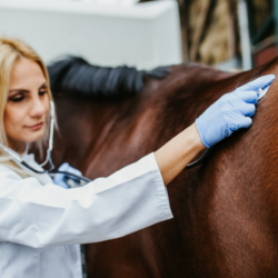
How is the condition diagnosed?
The diagnosis of navicular syndrome is based on a combination of clinical examination and medical imaging. The clinical examination includes static (resting) and dynamic (moving) observations. The plank test, although tricky to interpret, involves placing the horse’s foot on a plank and observing the pain reactions when the plank is lifted.
Local anaesthesia of the distal part of the foot can also be used to confirm the location of the pain. If locomotion improves after anaesthesia, this indicates that the pain originates in this area. However, this is not a definitive indication of podotrochlear syndrome.
Imaging techniques such as radiography and ultrasound are essential for observing bone structures and soft tissues. Radiography is used to visualise abnormalities such as bone fragments, radiolucent areas or changes in bone density.Ultrasound is used to examine soft tissues such as tendons and ligaments.
MRI (Magnetic Resonance Imaging) is the method of choice for a precise and comprehensive diagnosis. It allows a detailed assessment of the various structures of the foot and can detect lesions that cannot be seen on X-ray or ultrasound. However, this technique is expensive and may not be available everywhere.
What treatments are available?
Treatment for navicular syndrome aims to relieve pain and slow the progression of the disease. However, it cannot cure the condition. In cases of intense lameness, the horse should be rested to allow inflammation to subside and bone remodelling to take place.
Appropriate shoeing is essential to reduce stress on the podo-trochlear apparatus. Orthopaedic shoes such as egg-bar shoes or Napoleon shoes are often used to support the heels . This also makes it easier to start the foot. Collaboration between the vet and farrier is essential to adapt the shoeing to the horse’s specific needs.
Medicinal treatment may include non-steroidal anti-inflammatory drugs to reduce pain, corticosteroid injections for localised inflammation, and tiludronic acid infusions to combat bone reshaping. Other options, such as acupuncture and shock waves, may also be considered depending on the lesions observed. Physiotherapy and physiotherapy are other complementary therapies that can improve mobility, strengthen muscles and aid the horse’s recovery.
In serious cases, vets will consider neurectomy. This involves severing the palmar digital nerves to eliminate pain. However, this procedure is controversial because of the associated risks, such as loss of sensitivity and recurrence.
What are the natural alternatives?
As a complement to medical treatments, food supplements can help manage navicular syndrome. Harpagophytum-based supplements, for example, help to limit stiffness and improve joint flexibility. Biotin is also recommended to encourage good horn growth and strengthen the foot.
Plants with anti-inflammatory properties, such as harpagophytum and meadowsweet, can be used in regular courses. Other supplements, such as Artphyton, support blood circulation in the foot and joint function respectively . Phytotherapy can also stimulate the production of bone cells and prevent bone demineralisation.
Meadowsweet, rich in salicylates, is an effective anti-inflammatory plant. It is often used to treat pain and inflammation naturally. Bamboo promotes the reconstruction of destroyed tissue, while horsetail stimulates collagen synthesis. Blackcurrant has properties similar to those of cortisone, making it useful for reducing inflammation. Lithothamnion, rich in minerals, helps to remineralise bones.
Finally, compounds such as glucosamine and chondroitin, combined with trace elements such as copper, zinc and manganese, protect cartilage. They maintain its elasticity and improve joint suppleness. Hoof packing poultices can strengthen the sole and reduce foot sensitivity. This contributes to the horse’s general well-being.