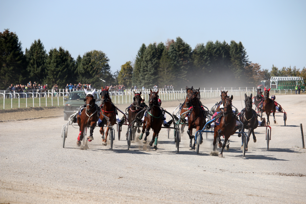
Tendinopathies represent a significant category of musculoskeletal injuries in sport horses, affecting their performance and general well-being. These pathologies are characterised by damage to the tendons, often caused by repetitive mechanical overload, trauma or biomechanical abnormalities. The most commonly affected tendons include the superficial flexor tendon of the finger (TFSD) and the deep flexor tendon of the finger (TFPD). Effective management of tendonitis requires a thorough understanding of the underlying pathophysiological mechanisms, as well as appropriate diagnostic, treatment and rehabilitation strategies.
What is tendonitis?
Tendonitis, inflammation of the collagen fibres in tendons, mainly affects active horses and is the second leading cause of career stoppage. Racehorses and sports horses are often prone to tendonitis of the superficial and deep flexor tendons.
The superficial (or “perforated”) finger flexor tendon lies just under the skin at the back of the barrel. The deep finger flexor tend on (or “perforator”) is covered by the perforator, attached to the carpus by the carpal tunnel and ends in the foot. The fetlock suspensory ligament (MIO3) divides into two branches halfway up the cannon, suspending the fetlock.
The tendons, which mature at an early age (2 years), do not develop an adaptive response to effort like the muscles, but have a high resistance to tension (up to 2,000 kg). If this resistance exceeds its limit, the tendon fibres are damaged.
The horse’s limbs, adapted to racing, contain few muscles but many tendons and ligaments. The large tendons have powerful accessory ligaments that automate movements and increase speed by saving energy. This automation complicates the management of tendonitis, as the horse has no control over the force exerted on its tendons, and the high stresses involved in movement lead to tendon injuries.
The superficial flexor tendon, an extension of the superficial flexor muscle of the finger, is attached to the forearm by the radial bridle and connects the pastern to the back of the forearm, passing under the fetlock.
The deep flexor tendon is an extension of the deep flexor muscle of the upper arm, attached to the carpus by the carpal tunnel, and descends to the foot, covered by the perforator. Unlike the perforated, it attaches to the third phalanx in the hoof, creating complementary tensions on the two flexor tendons.
What causes tendonitis?
Tendonitis in horses is mainly caused by work, combined with various risk factors. Poor posture (slouching, panties) increases the risk by putting excessive strain on the tendons. Unsuitable shoeing or trimming can place considerable stress on the tendons. Working on a ground that is too deep puts excessive strain on the tendons, causing the fetlock to hyperextend. The weight of the horse, particularly if it is overweight, puts extra strain on the tendons, as does intensive training. With age, the tendons deteriorate, making them more fragile. The use of incorrectly applied bands can also weaken the tendons.
Tendonitis of the deep flexor is often chronic and linked to repeated work, but can also result from trauma such as a laceration. Extrinsic factors, which can be influenced by the rider, include too deep a ground, overtraining and unsuitable shoeing. Intrinsic factors include poor legs, weight and age.
Acute tendonitis of the perforator tendon is rare compared to that of the superficial flexor tendon, which is often degenerative. Tendonitis is often caused by exertion, but trauma can also cause it. Factors favouring tendonitis include too deep a ground, excessive work, unsuitable shoeing, plumb defects, lameness of the other limb, excessive weight and age.
Tendinitis of the superficial flexor is mainly due to exertion, while that of the deep flexor is often linked to chronic fatigue or repeated shocks. Fatigue lesions heal with rest, while degenerative lesions require rest to relieve the horse. A lack of balance can lead to chronic tendonitis. Tendon injuries are common in sport horses, with the incidence varying according to the sport.
What are the symptoms?
How is it diagnosed?
If you suspect your horse has tendonitis, contact a vet as soon as possible for a diagnosis. Diagnosis begins with a three-stage clinical examination:
- Observation of the horse at a standstill to assess its balance and spot any deformities.
- Palpation to detect sensitive or hot areas.
- Examination on the move to observe locomotion on different surfaces and identify possible lameness.
After the clinical examination, an ultrasound scan is used to visualise the tendons, locate the tendonitis and assess its severity. The tendons most often affected are the deep (or “perforating”) flexor tendon, the superficial (or “perforated”) flexor tendon and the fetlock suspensory ligament.
To confirm involvement, truncal anaesthesia may be necessary to localise the lameness. Ultrasound is useful for identifying lesions, although limited for tendonitis in the hoof. X-rays can show calcifications in severe cases, but are often insufficient for a precise diagnosis.MRI is the test of choice for accurate diagnosis, despite its high cost (around €1,000). It is particularly recommended for chronic lameness.
Early signs of tendonitis include pain, swelling, stiffness, lameness, tenderness and thickening of the tendon. If these signs appear, rest your horse and contact a vet if symptoms persist. The examination will include observation of the legs, palpation of the affected area and, if necessary, an ultrasound scan. Ultrasound allows you to see the internal tissues and assess the extent of the inflammation.


