
Tularemia is a rare but serious infectious disease caused by the bacterium Francisella tularensis. Also known as “rabbit fever”, this zoonosis mainly affects lagomorphs and rodents, but can also affect a wide variety of other animals and humans.
What infectious agent is responsible?
Tularemia is a zoonosis caused by the bacterium Francisella tularensis. This small coccobacillus is Gram-negative, pleomorphic, aerobic, non-motile and non-spore-forming.
There are two main subspecies: F. tularensis subsp. tularensis (type A) and F. tularensis subsp. holarctica (type B). The tularensis subspecies is the most virulent. Fewer than ten bacterial cells cause infection. It is mainly found in North America, associated with lagomorphs. It is transmitted by ticks and biting flies.
On the other hand, subspecies holarctica is less virulent. It is found in Eurasia and North America. It is transmitted by aquatic rodents and arthropods, or by ingestion of contaminated water and food.
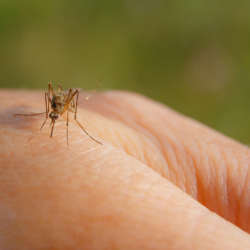
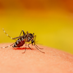
The main reservoirs of Francisella tularensis are lagomorphs and rodents. The bacterium can also be detected in various arthropods (ticks and mosquitoes), which play a crucial role as vectors. Ticks, particularly those of the Dermacentor genus, are the most competent vectors for the transmission of tularemia. The bacterium is extremely resistant to environmental stress, surviving for several months in water, soil and cadavers at low temperatures. It is sensitive to heat and to many disinfectants such as sodium hypochlorite, ethanol and formaldehyde.
Francisella tularensis is a potential biological threat and is on the list of Highly Pathogenic Microorganisms and Toxins (HPM). The bacterium mainly targets macrophages and dendritic cells, replicating in the cytoplasm of infected cells and inducing tissue necrosis. The bacterium’s virulence is linked to its ability to manipulate host immunity, evading initial detection and spreading via lymph nodes to various organs.
How does this disease manifest itself in animals?
Tularemia mainly affects lagomorphs (rabbits and hares) and rodents, but a wide variety of other mammals, birds, fish, amphibians and arthropods can also be infected. In Europe, outbreaks of tularemia are often sporadic, affecting mainly wild animals such as hares and rodents.
In susceptible animals, the disease is characterised by high fever, depression and often septicaemia. Infected animals show signs of severe lethargy followed by fatal septicaemia, with the disease progressing rapidly over two to ten days. At necropsy, lesions are often non-specific, with generalised congestion and enlargement of the liver and spleen.
Haematophagous ticks play a crucial role in maintaining and transmitting F. tularensis in the wild. Most domestic animals do not usually show clinical signs of tularemia, although they may develop specific antibodies following infection.
Among domestic animals, cats are more likely to develop clinical signs of tularemia than dogs.Cats transmit the disease to humans by biting or scratching. Dogs transmit the disease through close facial contact, ticks or scavenged carcasses. The disease in animals varies from acute to chronic forms. Acute forms progress to septicaemia and death within a few days. Chronic forms depend on the infecting dose.
How is it transmitted?
Tularemia is transmitted by several routes, mainly by direct contact with infected animals or by arthropod vectors such as ticks and mosquitoes. Transmission can occur via the skin, respiratory tract, conjunctiva or digestive tract.
By the cutaneous route, the bacteria can penetrate healthy skin through direct contact with contaminated animals, furs or organs, or through a wound (thorn, splinter) or a tick bite. This route is common among hunters, gamekeepers and foresters who regularly handle game.
By the respiratory route, tularemia can also be transmitted by inhalation of dust contaminated with the faeces of small mammals or cadavers. Such transmission is common in workplaces where soiled fodder, cereals or bedding are frequently handled.
Through the digestive tract, consumption of contaminated water or undercooked meat from infected animals is another route of transmission. This route of contamination is of particular concern in regions where drinking water resources are limited.
People at risk include professionals working in direct contact with wild rodents or small game, animal dealers, veterinary laboratory staff and taxidermists. Transmission can also occur through ingestion of contaminated food or water, bites or stings from infected arthropods, or inhalation of contaminated aerosols or dust.
Tularemia is highly contagious, but there is no direct person-to-person transmission. The bacteria can persist in the environment and in arthropod vectors, such as ticks, throughout their lives.
What are the symptoms of this infection in humans?
Tularemia manifests itself in two main forms in humans, depending on the route of entry. Local infection occurs after a bite or contact with infected animals. It is characterised by ulcers or abscesses and swollen lymph nodes. Generalised infection occurs after ingestion or inhalation of the infectious agent. It can lead to severe complications, such as damage to the lungs, digestive tract or brain. The disease begins after an incubation period of 3 to 15 days, with high fever, chills, flu-like symptoms, myalgias, arthralgias, headaches, and sometimes nausea, vomiting and diarrhoea.
The clinical forms of tularemia depend on the route of entry of the causal agent:
- The ulcero-ganglionic form (80-90% of cases): local inflammation at the point of penetration, progressing to necrotic ulceration and regional adenopathy.
- The lymph node form: regional adenopathy without skin involvement.
- The oculoganglionic form: conjunctivitis with regional adenopathy after ocular contact.
- The oropharyngeal form (especially in children): ingestion of contaminated food or water, stomatitis, ulcerative pharyngitis and cervical adenopathy.
- The pleuropulmonary form: inhalation of contaminated aerosols, dry cough, bronchiolitis, pleuropneumonia and respiratory distress.
- The typhoid or septicaemic form: high fever, headache, malaise, vomiting, diarrhoea and abdominal pain.
The lethality of infection by subspecies tularensis can reach 30% without treatment, while that of subspecies holarctica is less than 1%. The immune response, with specific IgM, IgA and IgG antibodies, is crucial for defence against this bacterium.
How is it diagnosed?
Diagnosing tularemia poses major challenges. Isolating the bacteria by direct culture remains difficult. Even in large quantities in lymph node pus, growth on culture medium is rare. Blood cultures are generally negative. To improve the sensitivity and speed of diagnosis, it is recommended that the sample be inoculated into a mouse or guinea pig. The spleen of the sacrificed animal is then subcultured.
Serodiagnosis is crucial for diagnosing tularemia. However, serological tests only become positive after two weeks of illness. Wright’s sero-agglutination method, using a suspension of killed Francisella tularensis, is commonly used. Serology can remain positive for several years. It is therefore necessary to observe a significant rise in antibody titres to diagnose an acute infection.
Biological samples for the bacteriology laboratory include serosities from the point of inoculation. Ocular or pharyngeal exudates, sputum and pleural fluid are also collected. The bacteria can be identified by direct immunofluorescence or immunohistochemistry, but these techniques are rare.
PCR (polymerase chain reaction) amplifies and detects the DNA of F. tularensis, offering early diagnosis in cases of strong clinical suspicion. This method reduces the risk of contamination associated with bacterial cultures. Isolation of the bacterium from clinical samples (skin lesions, lymph node punctures, blood, cerebrospinal fluid) remains possible. However, this requires high-security laboratories (P3).
What is the appropriate treatment?
Treatment of tularemia is based on appropriate antibiotic therapy, which should be administered as soon as possible after diagnosis to avoid severe complications. First-line antibiotics include aminoglycosides (gentamicin, streptomycin), fluoroquinolones (ciprofloxacin) and tetracyclines (doxycycline).
Aminoglycosides are effective in treating severe systemic forms of tularemia, while fluoroquinolones and tetracyclines are suitable for less severe forms or for per-oste treatment. Streptomycin and gentamicin are the antibiotics of choice, although there is no known acquired resistance for Francisella tularensis to date.
Moderate clinical forms require oral treatment with a fluoroquinolone (ciprofloxacin 500 mg twice daily or ofloxacin 400 mg twice daily) or doxycycline (100 mg twice daily) for 14 days. Severe forms may require a combination of two antibiotics, for example an aminoglycoside and a fluoroquinolone.
In the case of lymph node suppuration, surgical removal of the affected lymph node(s) may be necessary in addition to antibiotic treatment. The French Ministry of Health recommends hospitalisation for patients with severe or systemic clinical forms of tularemia.
A vaccine against tularemia is available in the United States, although it has not yet been approved for human or animal use in Europe. Hospitalisation of patients depends on the clinical severity and the subtype of F. tularensis responsible for the symptoms.
What preventive measures are available?
If tularemia is suspected in an animal, additional precautions include wearing protective goggles and wetting fur with a disinfectant before handling. Waste and cadavers must be transported in sealed, labelled bags or containers.
Some epidemiological data…
Tularemia is a notifiable disease in France, where it is relatively rare, with 80 to 100 cases reported each year. The disease is endemic throughout the northern hemisphere, with sporadic outbreaks in Europe, particularly in Scandinavia, Eastern Europe and Turkey.
In Belgium, only 14 cases were recorded between 1950 and 2017, including 11 between 2012 and 2017. The number of cases has increased in recent years, with five cases reported in 2017. In Europe, the notification rate in 2015 was 0.25 per 100,000 inhabitants, with 1,121 confirmed cases, mainly in Sweden, Finland, Norway, Hungary and the Czech Republic.
The subspecies of F. tularensis occupy specific geographical areas: subspecies tularensis predominates in North America, while subspecies holarctica extends throughout the northern hemisphere and appears in Tasmania and Australia. Subspecies mediasiatica is restricted to Central Asia.
Exposure factors to tularemia vary, including contact with wildlife, tick bites and contact with contaminated environments. Tularemia is a recognised occupational disease in livestock farmers, veterinarians, slaughterhouse workers, game wardens, forest rangers and laboratory staff.
In the laboratory, contamination can occur via the cutaneous, ocular, oral or respiratory route. F. tularensis cultures and infected animals are handled in level 3 biosafety laboratories.