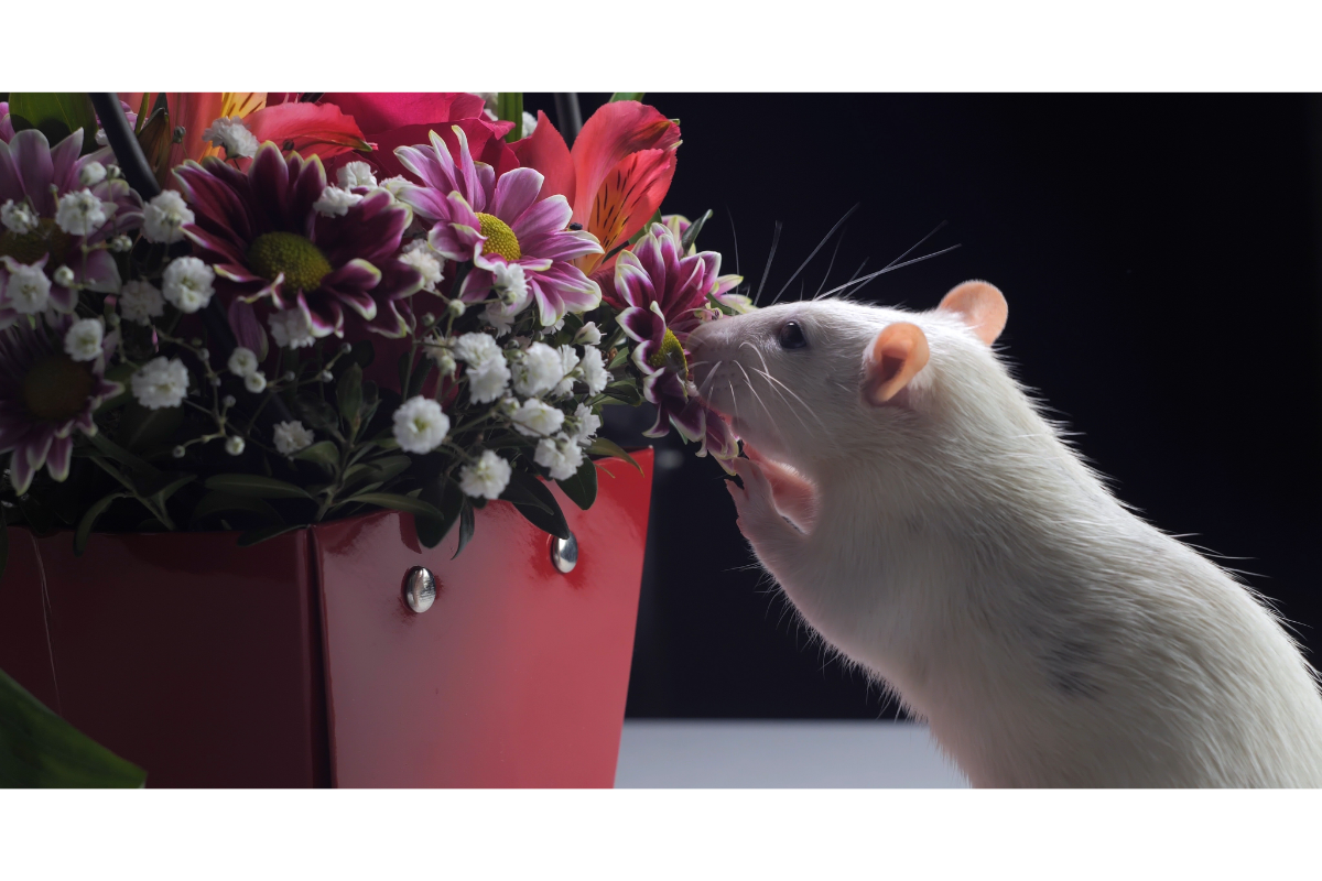
Lymphocytic choriomeningitis (LCM), also known as viral choriomeningitis, is a viral disease caused by a virus of the genus Lymphocytic choriomeningitis virus (LCMV). Although rare, this infection can have serious consequences for human health. What’s special about LCM is that it is often transmitted to humans by infected rodents. This makes it a zoonosis, i.e. a disease that can be transmitted from animals to humans.
What is the virus responsible for this disease?
Morphologically, VCML appears as a round, oval or pleomorphic virion, with a diameter varying between 110 and 130 nm. Its genome consists of single-stranded RNA in two segments. The virus affects a variety of hosts, including humans, mice, hamsters, guinea pigs, pigs, rats, monkeys, dogs, rabbits and chickens. The incubation period is estimated at between 8 and 13 days before the first meningeal symptoms appear.
Ribavirin has been shown to be effective in reducing the symptoms of the infection, while bleach and other common household disinfectants can inactivate VCML. It is also sensitive to UV rays and heat.
Outside its host, VCML is rapidly inactivated unless stored at -80°C. In the laboratory, the virus has been shown to retain its infectivity for at least 206 days in 0.85% saline containing 50% glycerine, at 4-10°C.
The lymphocytic choriomeningitis virus, as a model in immunology and virology, has led to major advances in our understanding of various aspects of human immunity and viral pathology. In particular, its study has shed light on areas such as immunological tolerance, antigenic presentation, restriction of the major histocompatibility complex, the role of T lymphocytes in viral elimination,T lymphocyte exhaustion and immune memory.
How does the disease affect animals?
The lymphocytic choriomeningitis virus infects a wide range of species. The main species that transmit the disease are the common mouse or grey mouse, as well as laboratory and domestic rodents such as mice, hamsters, guinea pigs, rats and squirrels. However, it is important to note that the virus can infect other species, such as dogs, cats and ruminants, without being transmitted to other individuals.
The geographical distribution of the lymphocytic choriomeningitis virus is worldwide. This means that it can be found in many regions around the world. However, the frequency of cases in pet rodents is not well documented. This makes it difficult to assess the extent of infection in this population.
Symptoms of infection with lymphocytic choriomeningitis virus vary from one individual to another. In many cases, the infection may be asymptomatic, meaning that the infected animal shows no clinical signs of the disease. However, in some cases, nervous symptoms may appear, such as coordination disorders, convulsions, paralysis and signs of respiratory distress. In the most serious cases, these symptoms can lead to the rapid death of the infected animal.
How is this virus transmitted?
Lymphocytic choriomeningitis infection in animals is transmitted in a variety of ways. When infection occurs in adults, it is generally transient. However, if it occurs in utero or around birth, it persists throughout the animal’s life. Infected animals excrete large quantities of the virus in various secretions and excretions, especially urine. Contamination can therefore occur through biting, contact with injured skin or mucous membranes, inhalation of aerosols or digestion.
In humans, most lymphocytic choriomeningitis infections occur through inhalation of dust or ingestion of food contaminated with the urine, faeces or other biological fluids of infected mice or hamsters. Transmission occurs mainly through biting, contact with the faeces or saliva of infected rodents, inhalation of contaminated dust aerosols during close contact with infected rodents, or more rarely via the digestive tract through consumption of contaminated food or water. The disease is considered rare, with exceptional cases reported in France.
The incubation period is approximately 8 to 13 days before the first meningeal symptoms appear. There is no evidence of human-to-human transmission, with the exception of vertical transmission from mother to foetus during pregnancy and organ transplants from infected donors.
The main reservoir of the virus is the common mouse, although the Syrian hamster can also be a reservoir. The virus is spread mainly by contact with the secretions or excretions of contaminated rodents, with a possible presence in various vectors such as fleas, midges, mosquitoes, ticks and cockroaches, although their role in transmission is unlikely.
How does choriomeningitis manifest itself in humans?
The incubation period for lymphocytic choriomeningitis is generally 1 to 2 weeks. Most infected people have no or mild symptoms. However, in some cases, symptoms may appear after 1 to 2 weeks of infection.
The most common symptoms include a flu-like illness with fever, chills, general malaise, weakness, muscle aches (particularly in the lower back), pain behind the eyes, sensitivity to light, loss of appetite, nausea and dizziness. Sore throats are less common.
After a period of 5 days to 3 weeks, most people feel better for 1 or 2 days. However, in some people, the condition may deteriorate again, with a recurrence of fever, headache and possibly a skin rash. Swelling of the joints in the fingers and hands may also occur. In some cases, the infection can spread to the salivary glands and testicles.
In a few individuals, an infection of the meninges(meningitis) may occur, manifested by a stiff neck that makes it difficult or impossible to move the chin towards the chest. In rare cases, a brain infection(encephalitis) may develop, leading to symptoms such as paralysis or other brain dysfunctions.
In pregnant women, the infection can have serious consequences for the foetus, leading to problems such as hydrocephalus, chorioretinitis and intellectual impairment. Complications can include blurred vision, eye pain, sensitivity to light and even blindness. If the infection occurs during the first trimester of pregnancy, the foetus may even die.
How is the disease diagnosed?
Looking for symptoms is crucial for monitoring lymphocytic choriomeningitis. The diagnosis is confirmed by various means, including serology,ELISA, RT-PCR, Western blot, immunohistochemical staining, neutralisation tests,immunofluorescence and viral culture using blood or cerebrospinal fluid. It should be noted that not all of these methods are available in all countries. The complement fixation test, although widely used, is now considered to be insensitive. Its use is no longer recommended.
Diagnosis of lymphocytic choriomeningitis usually involves a lumbar puncture to collect a sample of cerebrospinal fluid. This fluid is analysed for the presence of a virus, using RT-PCR to detect viral RNA, or serology to detect antibodies to the virus.
In individuals with symptoms suggestive of meningitis or brain infection who have been exposed to rodents, lymphocytic choriomeningitis is suspected. Doctors may carry out blood tests to look for the presence of antibodies directed against the virus. This helps to confirm the diagnosis.
How is it treated?
The treatment of lymphocytic choriomeningitis is based primarily on supportive care designed to alleviate symptoms and maintain the patient’s vital functions. This care includes management of symptoms such as fever, headache, muscle pain and other clinical manifestations associated with the infection. It is essential to help the patient through the acute period of illness and to promote recovery.
In cases where the illness is particularly serious, such as meningitis or a brain infection, hospitalisation is often necessary. While in hospital, patients may receive antiviral treatment, in particular ribavirin, which has been shown to be effective in vitro against the virus responsible for lymphocytic choriomeningitis. The administration of ribavirin can help to reduce the viral load and alleviate symptoms, thus offering relief to the patient and encouraging the disease to progress more smoothly.
In some cases, corticosteroids may also be considered. Corticosteroids have anti-inflammatory properties and can help to reduce the inflammation associated with infection, particularly in cases where inflammatory complications arise, such as meningitis.
The treatment of lymphocytic choriomeningitis is based on a multidisciplinary approach designed to relieve symptoms, limit complications and promote patient recovery. This includes supportive care, close monitoring of symptoms and, in some cases, the administration of specific drugs such as ribavirin and corticosteroids.
What can be done to prevent the disease?
We recommend sourcing animals from farms that regularly screen for lymphocytic choriomeningitis infection. It is also important to prevent any direct or indirect contact between farmed rodents and wild rodents, particularly mice.
As far as general hygiene on the farm is concerned, it is essential to control the presence of rodents by avoiding attracting them with food deposits or cluttered premises, and by carrying out regular rodent extermination. It is also essential to limit exposure to dust when cleaning the premises by airing them out and using a hoover, while regularly cleaning and disinfecting the premises, equipment and cages.
Employees must be informed of the risks associated with lymphocytic choriomeningitis, and of the collective and individual preventive measures to be adopted. This includes handling and restraining rodents, for which appropriate resources such as drinking water, soap and personal protective equipment must be provided. In addition, in the event of an animal disease, it is imperative to identify the source of contamination, reinforce hygiene and disinfection measures, and reduce possible sources of contamination.
To prevent infection, ventilate enclosed spaces where mice have been present before cleaning. Moisten surfaces with a 10% bleach solution before sweeping or cleaning. Avoid stirring up dust, and seal up openings through which rodents can enter homes. Store food in containers that are inaccessible to rodents. Finally, eliminate potential nesting sites around homes.
What is the status of choriomeningitis?
Lymphocytic choriomeningitis, although present in animals, is not considered to be a widely contagious disease. Unlike other highly infectious animal diseases, it is not generally transmitted significantly from one animal to another. However, its prevalence and impact can vary according to animal populations, husbandry practices and environmental conditions.
Although lymphocytic choriomeningitis can affect humans, it is not a notifiable disease. This means that cases of the disease are not systematically reported to the public health authorities. However, regular surveillance and appropriate management of detected cases are essential to assess and contain any potential risk to public health.
At present, lymphocytic choriomeningitis is not listed in the official tables of occupational diseases. This means that there is no official recognition of this disease as being directly linked to specific occupational activities. However, this does not mean that people exposed to this disease in the course of their work cannot benefit from specific protection and measures.
The virus responsible for lymphocytic choriomeningitis is classified into danger groups according to its virulence and infectious potential. Neurotropic strains, which tend to infect the nervous system, are classified in hazard group 3. This indicates a high risk to human health in the event of exposure. The other strains, although less dangerous, are classified in hazard group 2. This still highlights the need to take appropriate precautions when handling and studying them, in accordance with current regulations.


