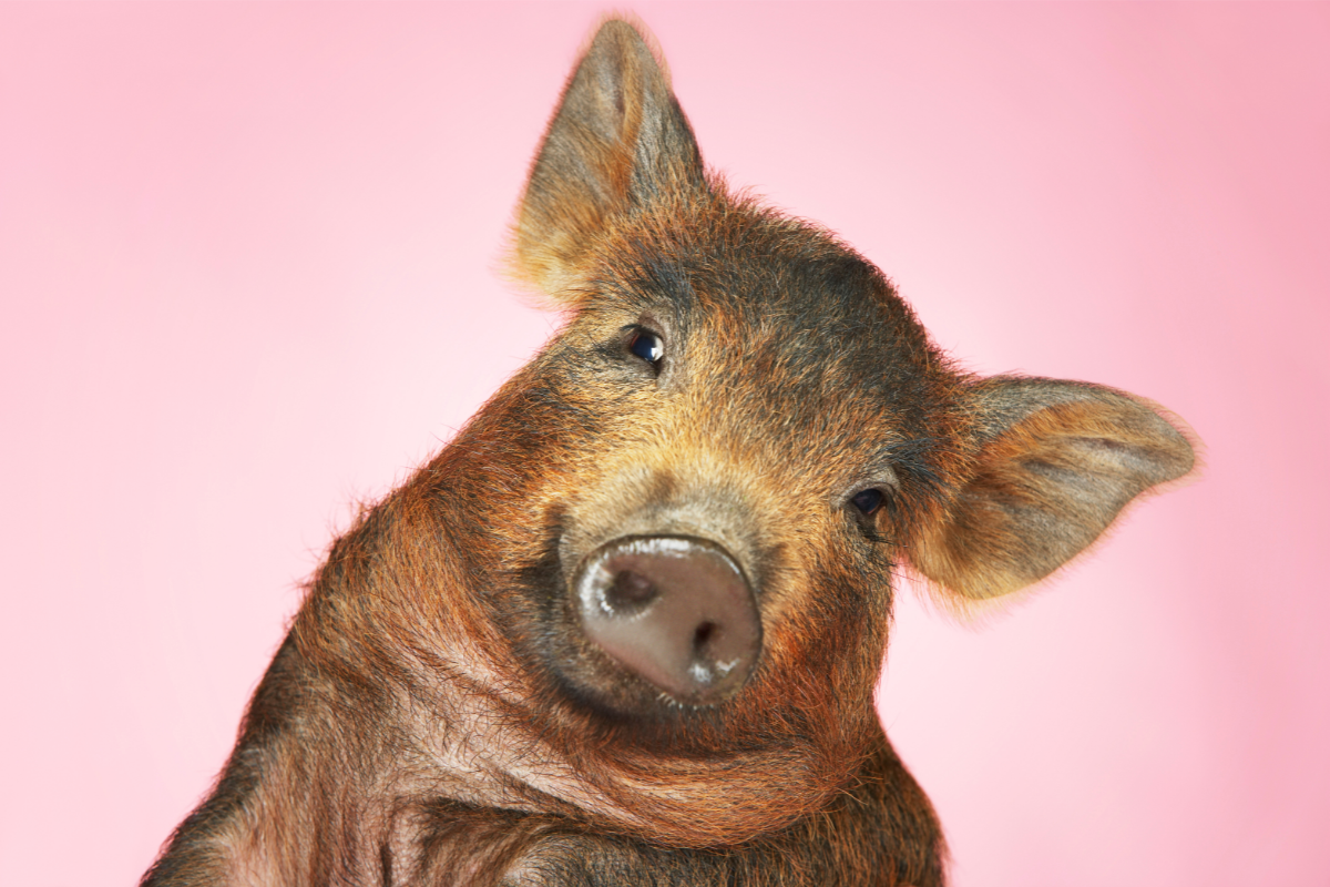
Porcine erysipelas is an infectious disease caused by the bacterium Erysipelothrix rhusiopathiae. This zoonosis, transmissible from animals to humans, is a significant concern for workers in close contact with pigs, such as breeders, butchers and vets.
What is the bacterium responsible?
Porcine rouget fever is a bacterial disease that mainly affects pigs, but occasionally also lambs, calves and humans. The bacillus Erysipelothrix rhusiopathiae, an unbranched rod-shaped Gram-positive bacterium, causes this zoonosis. Scientists initially identified E. rhusiopathiae as an animal pathogen responsible for erysipelas. In humans, this zoonosis is known as Rosenbach’ s erysipelas or rouget du porc, and is recognised as an occupational disease in France.
Turkeys and pigs are the species most frequently affected. However, cases have also been observed in other birds, fish and reptiles. Human infections with E. rhusiopathiae often present in an attenuated cutaneous form known as erysipelotrichosis.
Morphologically, E. rhusiopathiae is an immobile bacillus, very thin (2 µm by 0.2 to 0.4 µm), anaerobic or facultative aerobic, capable of forming filaments. In culture, this bacterium grows between 5 and 44°C, at neutral pH, preferring a CO2-enriched atmosphere. Colonies are very small, α-hemolytic, catalase negative, oxidase negative and H2S positive. Blood agar can be made selective by adding azide, crystal violet and novobiocin.
Identification is possible using microarrays such as API Coryne. There are around twenty polysaccharide capsular serovars. When inoculated intraperitoneally into mice, the bacillus causes rapidly fatal septicaemia. Heating to 70°C for 5 to 10 minutes, as well as phenol or sodium hydroxide solutions, can destroy this bacterium.
Pathogenicity factors include a heat-labile capsule and enzymes such as hyaluronidase and neuraminidase. E. rhusiopathiae survives for several weeks in the environment and is resistant to desiccation, cold and freezing.
How does red mullet affect pigs?
Many species can be infected by the rouget bacillus. The farm animals most affected are pigs, sheep and poultry, but various fish and shellfish from contaminated waters can also carry the germs without showing any signs of disease.
Red mullet is found worldwide. Transmission occurs via the digestive tract or the skin (foot lesions, castration wounds, umbilical wounds). The outside environment (farm floor and grazing land) is contaminated by the faeces of sick animals or healthy carriers. All tissues (meat) and faeces from sick animals are contaminated.
Symptoms vary according to species:
- Pigs: acute form (septicaemia, fever, prostration, purplish rash, death in 2-3 days without treatment), localised forms (arthritis, cardiac damage, abortion).
- Lambs: mainly arthritis.
- Birds: septicaemia progressing to death in 24-48 hours (20-50% of birds).
- Other species: septicaemia, cardiac damage, arthritis, subcutaneous abscesses (marine mammals).
In pigs, there are three forms of the disease:
- Superacute form: the most severe, with fever, bluish-reddish skin signs, death within a few hours.
- Acute form: moderately severe, with lesions that can progress to necrosis of the limbs, ears or tail, followed by death.
- Chronic form: benign, causing joint inflammation.
Erysipelothrix rhusiopathiae infects a wide range of animals, with or without causing clinical disease. The main reservoir is the pig (30-40% of healthy pigs). Many other species are also carriers, including sheep, ruminants, fish, shellfish, birds, poultry and rodents. Contamination of the external environment occurs through soil and water.
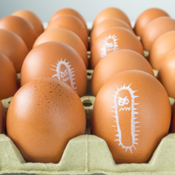
In poultry, the bacterium has been isolated from numerous avian species, with outbreaks reported in almost all poultry species, mainly turkeys and laying hens.
How is it transmitted?
Red mullet is mainly transmitted through the skin, by accidental inoculation (puncture) or by contamination of a pre-existing wound. There is no human-to-human transmission.
The frequency of cases of red mullet remains poorly documented in mainland France and the French overseas departments. Occupations at risk include working in direct contact with infected animals or in contaminated environments (bedding, farm premises, transport vehicles), as well as handling contaminated meat, offal, viscera, bones and bones. The occupational categories most at risk are slaughterhouse staff, butchers, rendering staff, naturalists, fishmongers, scalers, fishermen, veterinary surgeons, livestock farmers and veterinary laboratory staff.
Despite the strong resistance of the human species to E. rhusiopathiae, cases of infection mainly occur in specific professional environments. Medical analysis laboratories have not reported any infections. Documented cases have mainly come from people working in abattoirs, butchers’ shops, fishmongers, food industries, pig farms, and more rarely in kitchens and among veterinary surgeons. Currently, between 5 and 10 cases a year are recognised as an occupational disease.
Infection by E. rhusiopathiae generally occurs through skin wounds, often on the hands, in contact with infectious material or contaminated instruments. The veterinary and catering sectors are particularly concerned, where wounds caused by bone splinters, fish bones or scales can facilitate transmission. Asymptomatic carriers, although healthy, spread the bacteria through their excrement, contaminating the environment. The bacteria then enter humans or other animals through a wound or skin erosion. Although contamination is common, it does not always lead to infection.
The Erysipelothrix rhusiopathiae bacterium is ubiquitous, present everywhere in the environment. The bacterium’s strong resistance to a variety of environmental conditions, including damp soil and organic matter, contributes to its persistence and the spread of red mullet.
What does this infection look like in humans?
Infection by Erysipelothrix rhusiopathiae mainly manifests itself in a benign cutaneous form known as Baker-Rosenbach erysipeloid. It appears 24 to 48 hours after inoculation, in the form of a hard, slightly raised , purplish-red patch, accompanied by itching and burning. Healing generally takes 2 to 4 weeks. If left untreated, complications may include arthritis, heart disease or septicaemia.
Generalised forms, although rare, are serious and can lead to heart damage, septicaemia and generalised skin rash. Infection often occurs following skin wounds, especially on the hands, in contact with infectious material or contaminated instruments.
Typical lesions are purplish erythematous patches with pain, burning and pruritus at the site of inoculation. If left untreated or in immunocompromised patients, the infection may progress to arthritis, septicaemia or endocarditis. These serious complications are rare, except in immunocompromised patients.
Erysipelas is often an occupational disease, characterised by an erythematous oedema with well-defined, raised borders, usually localised on the backs of the hands and/or fingers. Palms, forearms, arms, face and legs are rarely involved. Vesicular, bullous and erosive lesions may also be present. The lesion may be asymptomatic or accompanied by mild pruritus, pain and fever.
Incubation lasts from 12 to 48 hours, and up to 10 days. Symptoms include a purplish erythematous plaque with pain, burning and pruritus at the site of inoculation. Resolution occurs in 2 to 4 weeks. In the absence of treatment or in immunocompromised patients, complications such as arthritis, septicaemia or endocarditis may occur. Rarely, the infection spreads to the lymph nodes, making them painful and swollen. Fever and other systemic symptoms are rare, except in cases of septicaemia, which is fortunately very rare.
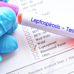
How is it diagnosed?
Diagnosis of Erysipelothrix rhusiopathiae infection is primarily clinical in cases of local infection. The patient typically presents with pruritus, pain, burning and redness of the skin at the site of inoculation. The lesion is often a purplish erythematous plaque (erysipeloid) with well-defined borders, usually appearing 24 to 48 hours after inoculation. This clinical presentation is sufficient to diagnose an erysipeloid in the majority of cases.
Infection frequently occurs through skin wounds, often on the hands, in contact with infectious material or contaminated instruments. If left untreated, or in immunocompromised patients, the infection can progress to serious complications such as arthritis, septicaemia or endocarditis.
Additional tests may be required to confirm the diagnosis, or in the case of disseminated forms. Isolating the germ is difficult, but is possible using a skin biopsy. In cases of suspected septicaemia, doctors recommend blood cultures, and in cases of joint involvement, they recommend culture of the joint fluid. Laboratories perform the culture on blood agar under CO2 and identify the samples by mass spectrometry. PCR detection is also possible, offering a rapid and accurate alternative for identifying the presence of the bacteria.
In general, the doctor makes the diagnosis of erysipelas based on the patient’s story and observation of the characteristic skin rash. However, in cases of doubt or atypical presentation, a skin sample (biopsy) is taken for further examination. This precaution, although rarely necessary, confirms the diagnosis and excludes other dermatological conditions.
Although the diagnosis of erysipeloid is based primarily on clinical examination, laboratory techniques such as culture on blood agar under CO2, identification by mass spectrometry and PCR confirm the presence of E. rhusiopathiae in complex or disseminated cases. These diagnostic tools are essential for appropriate management, especially in patients at risk of serious complications.
How is it treated?
In general,erysipeloides disappear spontaneously without any specific treatment. However, treatment with antibiotics can speed up healing and shorten the duration of the disease. The treatment of choice is a single dose of benzathine benzylpenicillin administered by intramuscular injection. Alternatively, a five-day to one-week course of oral penicillin or intramuscular benzylpenicillin procaine is also effective.
For patients allergic to penicillin, alternatives such as erythromycin or doxycycline can be used. It is crucial to note that Erysipelothrix rhusiopathiae is intrinsically resistant to vancomycin. However, this bacterium is highly sensitive to β-lactams, particularly penicillin G. Macrolides are used in cases of penicillin allergy and offer a good therapeutic alternative.
E. rhusiopathiae shows resistance to aminoglycosides, colistin and nalidixic acid, but remains susceptible to second-generation quinolones. It also shows resistance to novobiocin, similar to Gram catalase shells, due to the absence of a respiratory chain.
Standard treatment for a local infection involves the use of β-lactam (penicillin A or G) for one week. In the case of endocarditis, a serious form of the infection, treatment extends over a month to completely eradicate the bacteria. Patients allergic to β-lactams should use doxycycline or a macrolide as alternatives.
Appropriate treatment remains essential to prevent potential complications such as arthritis, septicaemia or endocarditis, particularly in immunocompromised patients. Although the infection may resolve spontaneously, the administration of antibiotics remains standard practice to minimise risk and speed recovery.
Although erysipelas can disappear without intervention, doctors recommend treatment with antibiotics to speed up healing and prevent complications. Alternatives such as erythromycin and doxycycline are suitable for patients allergic to penicillin.
What can be done to prevent it?
Rouget du porc is not a contagious animal disease, nor is it a notifiable human disease. However, the authorities consider this disease to be compensable at work (table no. 51 in the agricultural scheme, no. 88 in the general scheme). The worker or his heirs must make the declaration. The French Labour Code classifies Erysipelothrix rhusiopathiae in hazard group 2.
Collective preventive measures include :
- Vaccination of pigs, preferably targeting breeding males. However, the vaccine does not protect against chronic forms of the disease and may promote arthritis.
- General farm hygiene.
- Training and information for exposed personnel.
- Putting in place resources to guarantee hygiene, any necessary treatment, clothing and PPE (personal protective equipment) for exposed personnel.
- Reinforcing hygiene in the event of an animal disease.
To reduce the individual risk of contamination, compliance with hygiene protocols is essential:
- Reducing exposure: wearing gloves and boots when handling dead animals, giving birth, and when exposed to droppings. Droppings should not be cleaned with high-pressure water jets.
- Observe simple hygiene rules: wash hands, do not eat, drink or smoke in the workplace, and change clothes after work.
- No sick animals should be slaughtered for consumption.
General preventive measures include :
- Vaccination of pigs, mainly breeding stock.
- General farm hygiene, with cleaning and disinfection of premises and equipment.
- Storage of animal waste and carcasses in appropriate containers.
Employee training and information are crucial to raising awareness of the risks. In the event of an animal disease, it is essential to improve hygiene on the farm, isolate sick animals and restrict access to essential professionals. Washing and disinfecting contaminated sites and reusable service equipment must be rigorously applied.
History and epidemiology
In the nineteenth century, red mullet, then known as mal rouge, caused havoc in Europe and the United States. In 1879, the United States lost around 900,000 pigs, a financial loss estimated at 100 million francs at the time. In 1877, Achille Maucuer, a veterinary surgeon from Bollène in the Vaucluse region, concerned about the ravages caused by red mullet, drew Louis Pasteur ‘s attention to the disease. However, due to a lack of funds, Pasteur did not really begin his research until 1881. That same year, Louis Thuillier, under Pasteur’s supervision, isolated the bacterium responsible for rouget, which he named Erysipelothrix rhusiopathiae.
In 1883, after numerous vaccination tests in France, Pasteur announced that he had developed a vaccine against red mullet in pigs. At the time, the human form of the disease was not yet known. In correspondence with Maucuer in 1883, Pasteur expressed his astonishment at a potential case of human infection and suggested an investigation.
Today, although rare, the human form of swine mullet has been documented. In 1877, Maucuer alerted Pasteur to the red disease, which killed more than 20,000 pigs in the Rhône valley. Between 1876 and 1880, Robert Koch isolated the bacillus from a mouse injected with putrefied blood. In 1882, Pasteur asked Thuillier to study the disease in the Vienne region. Thuillier quickly identified a new microbe in the blood of dead pigs. This microbe, initially called Bacillus insidiosus, was given its current name in 1909.
In November 1882, Pasteur, accompanied by Thuillier and Loir, travelled to Bollène to examine the infected pigs. After attenuating the virulence of the microbe in rabbits, Pasteur obtained an effective vaccine for piglets. In 1883, the vaccine was welcomed by the local authorities. The Bollène town council expressed its gratitude to Pasteur.
Today, the human form of red mullet remains rare. Slaughterhouses, butchers, fishmongers, food industries and pig farms are the main sources of cases. The authorities recognise between 5 and 10 cases each year as an occupational disease. Before 1985, around 50 cases were recorded.