
Mediterranean spotted fever (MSF), also known as spotted fever, is an infectious disease caused by the bacterium Rickettsia conorii. It is transmitted by ticks of the genus Rhipicephalus. Although it is mainly found in Mediterranean regions, it can also be found in other parts of the world where the vector ticks are present. The disease is characterised by fever, muscle aches, headaches and a maculopapular rash, often mistaken for measles. Although rarely fatal, FBM can lead to severe complications if not treated promptly.
What is the pathogen?
Mediterranean spotted fever (MRF ) is a tick-borne disease caused by the bacterium Rickettsia conorii conori. It is transmitted mainly by the dog tick, Rhipicephalus sanguineus. Although it is widely present in Mediterranean regions, its eco-epidemiology remains poorly understood, particularly with regard to its original reservoir and factors of emergence. FBM is spreading geographically, and is becoming increasingly serious, making it one of the world’s emerging or re-emerging diseases.
Historically considered relatively benign, with a mortality rate of less than 2%, FBM has seen an increase in severe forms since the 1990s, with mortality rates of more than 3% and even as high as 6%. Rickettsia conorii is an obligate intracellular Gram-negative bacterium, mainly found around the Mediterranean basin, including southern France, where it was once responsible for ” typhus of the grape harvest “.
Although not found in the Americas, Australia and the Pacific, imported cases are possible after returning from endemic regions. Rickettsiae mainly infect arthropods and measure around 300 nm. They are classified as alpha-proteobacteria. Blood-sucking arthropods often inoculate humans with these bacteria, leading to various diseases, such as rickettsial disease.
For a long time, these bacteria were considered to be intermediate between bacteria and viruses, due to their intracellular nature and small size. Rickettsiae penetrate cells where they multiply by scissiparity, causing multifocal infections, particularly of the vascular endothelium.
The pathogenesis of FBM involves a multifocal infection with a disseminated vasculitis, explaining the various clinical manifestations, such as cutaneous eruptions and cardiac, muscular, renal and neurological complications.
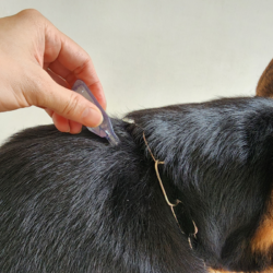
How does the disease affect animals?
In dogs, the symptoms of Rickettsia conorii infection are generally absent or minimal. The infecting bite is often caused by the immature forms of the tick (larva, nymph), often going unnoticed because of its small size and discretion. The tick must remain in contact with the skin for 10 to 20 hours to transmit the infection.
Dogs do not act as reservoir hosts for the bacterium, which is incapable of multiplying in animals, causing only a transient fever. Instead, the dog acts as a vector, carrying infected ticks to humans. The ticks themselves act as a reservoir, transmitting the infection to their offspring via the ovaries.
Although wild rabbits are considered the likely reservoir of Rickettsia conorii, other potential reservoirs, such as hedgehogs or other small rodents, are also possible. The disappearance of rabbits due to myxomatosis in France in 1952 led to a drop in cases of infection, followed by an increase after the reappearance of wild rabbits in 1967.
How is the disease transmitted?
Mediterranean spotted fever is transmitted by the bite of a brown dog tick(Rhipicephalus sanguineus). Infected dogs or tick carriers do not transmit the disease directly to humans, but their presence increases the risk of infection.
In south-eastern France, sporadic cases of Mediterranean spotted fever have been linked to the presence of ticks. Occupational activities at increased risk include those involving tick bites, such as working in places regularly frequented by dogs, such as kennels and forestry work.
The brown dog tick(Rhipicephalus sanguineus) is the main vector of this disease. Although the probability of being bitten is low, especially in humans, this tick is more aggressive when the temperature rises. In the south of France, where these ticks are active between May and October, the majority of human cases are diagnosed in July and August.
After an incubation period of around a week, symptoms of Mediterranean spotted fever can appear gradually, resembling a flu-like condition, or suddenly, with chills, fatigue, headaches, aches and pains and a high fever (39-39.5°C) within a few hours. Infection generally occurs in summer when ticks bite in scrubland and gardens, particularly along the Mediterranean coast.
In southern France, the number of annual cases of spotted fever has fallen sharply since myxomatosis reduced the population of rabbits, which are hosts to the causative bacterium, Rickettsia conorii. A black spot may appear at the site of the bite, which may eventually ulcerate.
What are the symptoms of spotted fever?
Symptoms of Mediterranean spotted fever include a sudden onset of high fever, accompanied by muscle aches and headaches. These symptoms are often associated with the presence of a ganglion and, more frequently, a black crusty spot at the site of the tick bite. Subsequently, a rash appears all over the body, with the exception of the face, including on the palms and soles of the feet.
The incubation period lasts an average of a week (3 to 16 days). The onset of symptoms is sudden, with a clinical picture resembling an influenza-like illness, including a fever of 39°C, intense headaches, photophobia and muscle and joint pains. In more than half the cases, the tick bite site is found in the form of a black spot.
This painless, crusted lesion, 3 to 5 mm in diameter, is surrounded by an inflammatory halo measuring 2 to 3 cm. It may be accompanied by adenopathy and lasts about a week before healing. Ocular inoculation can lead to conjunctivitis.
The skin eruption begins as a morbilliform exanthema on the trunk, then progresses to the limbs, changing from a macular to a papular form, and finally giving a pimply appearance. It may be associated with punctiform skin haemorrhages known as petechiae, and in severe cases, extensive purpura.
Although the course of the disease is generally benign, visceral complications can occur, such as digestive, neurological, cardiovascular and pulmonary damage, accounting for 5-6% of cases. Risk factors such as alcoholism, diabetes, G6PD deficiency, advanced age, delayed treatment or inappropriate antibiotic therapy can increase the risk of complications.
How is this disease diagnosed?
The diagnosis of Mediterranean spotted fever is suspected in the presence of symptoms suggestive of “summer flu”, especially in endemic regions or in individuals returning from trips to these areas. The presence of a dog in the vicinity, a cutaneous rash with a delayed onset and the presence of the characteristic black spot help to guide the diagnosis.
Various serological techniques are used to confirm the diagnosis, such as the classic Weil-Felix test (based on beef protein agglutination), the ELISA test or the immunofluorescence method. In addition, direct visualisation of rickettsiae on a biopsy of the inoculated eschar provides confirmation.
Molecular detection of rickettsia by PCR is another diagnostic method, although its availability may vary depending on the laboratory’s technical facilities. Clinical diagnosis is often easier in endemic areas, but outside these areas there may be confusion with influenza.
Because of cross-immunity, laboratory results may simply indicate “spotted group fever” following agglutination with Proteus X2. However, molecular diagnosis can be performed on serum or skin biopsy using specific probes for real-time PCR (RT-PCR) or even sequencing with target genes such as ompA, ompB, and gltA.
These genomic techniques make it possible to distinguish several subspecies of R. conorii, including R. conorii conorii (agent of Mediterranean spotted fever), R. conorii israelensis (agent of Israeli spotted fever), R. conorii caspia (agent of Astrakhan fever), and R. conorii indica (agent of Indian tick typhus).
How is it treated?
The treatment of Mediterranean spotted fever is based mainly on the use of antibiotics, in particular tetracyclines and macrolides. These drugs are effective against bacterial infections. These antibiotics are generally administered orally over a period of about a week. In some cases, treatment may be extended to two days after the fever has disappeared to ensure complete elimination of the infection and prevent relapses.
It is crucial to start antibiotic treatment as soon as possible after the disease has been diagnosed. This will prevent the infection spreading through the body and prevent the development of serious complications. Antibiotics work by killing the bacteria responsible for the infection or inhibiting their growth. In this way, they enable the immune system to fight the infection more effectively.
Close monitoring of the patient’s clinical progress is essential during treatment. If signs of complications or deterioration in health appear, adjustments may be necessary, including extending the duration of treatment or changing the antibiotic.
It should be noted that MBF, although generally considered to be a benign disease, can lead to serious complications, particularly in patients with severe forms of the disease. It is therefore essential that patients follow their doctor’s recommendations carefully. They must complete the prescribed course of antibiotics to ensure a full recovery and avoid long-term complications.