
Alveolar echinococcosis, a potentially fatal parasitic disease, is attracting increasing interest due to its complexity and rarity. Caused by the worm Echinococcus multilocularis, the condition mainly affects people living in close contact with reservoir hosts, such as foxes and dogs, in regions where the parasite is endemic. Although relatively rare, alveolar echinococcosis presents a diagnostic challenge due to its insidious symptomatology and the need for a multidisciplinary approach for effective management.
What is the pathogen?
Alveolar echinococcosis is caused by the larva of a flatworm of the Taeniidae family: Echinococcus multilocularis. The parasite’s life cycle involves three main compartments:
- the definitive hosts (mainly carnivores such as foxes, and to a lesser extent dogs and cats),
- intermediate hosts (rodents)
- and the environment.
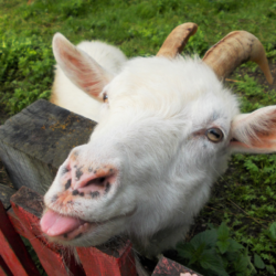
Carnivores are the main reservoirs of the adult echinococcus worm.
The development cycle is as follows:
- The adult worms reside in the intestines of the definitive hosts (mainly foxes), where they produce eggs. The eggs are excreted into the environment in the faeces.
- Intermediate hosts (often rodents) accidentally ingest parasite eggs by eating plants contaminated with excrement.
- The ingested eggs hatch into echinococcus larvae(metacestodes) which develop in the liver or lungs of the intermediate hosts.
- When carnivores (foxes, dogs or cats) predate infected rodents, the parasite’s life cycle ends. The carnivores ingest the protoscolices present in the tissues of the rodents, where they rapidly develop into adult worms.
Dogs and cats can be contaminated by Echinococcus multilocularis but play a minor role in the epidemiological cycle of the parasite. Echinococcus eggs are highly resistant and can survive in the environment for over a year. Even intensive washing does not guarantee total elimination of the parasite’s eggs, and only cooking fruit and vegetables at over 60°C can eliminate the risk of contamination. Domestic freezing does not inactivate parasite eggs.
What are the symptoms in animals?
Echinococcus multilocularis can infect various species, particularly carnivores such as foxes, dogs and sometimes cats. Carnivores play a crucial role in the parasite’s life cycle as definitive hosts. They harbour adult worms in their intestines and release parasite eggs into the environment via their faeces. In addition, wild rodents, particularly voles, act as intermediate hosts in the parasite cycle, harbouring echinococcus larvae and thus contributing to the spread of the disease.
The geographical distribution of Echinococcus multilocularis is restricted to the northern hemisphere, mainly in cold climate regions. In Europe, cases of infection are most frequent in northern and eastern countries such as Switzerland,Germany, Belgium and Italy, as well as in certain regions of France. In France, outbreaks are concentrated mainly in the north-east and the Massif Central, where environmental conditions favour the parasite’s survival and transmission cycle.
Symptoms of Echinococcus multilocularis infection are often not apparent in animals. In foxes, the disease generally remains asymptomatic, making detection of infection difficult without in-depth epidemiological studies. In dogs, symptoms may include digestive signs such as diarrhoea and coprophagia, although in most cases the presence of adult echinococci in the intestine does not lead to obvious clinical manifestations. It is therefore essential to carry out faecal analyses to detect the presence of the parasite in pets.
How is this parasite transmitted?
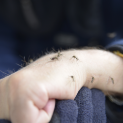
Echinococcus multilocularis is transmitted mainly via the digestive tract. Carnivores contract the parasite by eating small rodents infected with Echinococcus multilocularis. Once infected, carnivores house the worm in their small intestine and excrete the microscopic eggs of the parasite in their faeces, which adhere firmly to plants and soil, being highly resistant to environmental conditions.
Carnivores act as definitive hosts, harbouring the mature tapeworm in their intestines after ingesting the viscera of intermediate hosts containing the parasite’s larvae. Rodents contribute to transmission by ingesting food or water contaminated with the excrement of carnivores, thus containing eggs of the parasite.
There are several genotypes of Echinococcus granulosus, some with specific preferences in terms of intermediate hosts. Although alveolar echinococcosis generally arises from a cycle involving wild animals such as foxes and small mammals, domestic dogs and cats can also serve as definitive hosts for the parasite.
In humans, transmission is mainly oral. Individuals accidentally ingest parasite eggs present on contaminated plants, such as vegetables, mushrooms or wild berries, or by bringing to the mouth hands contaminated by eggs present on the fur of carrier animals such as dogs and cats. Humans thus act as accidental intermediate hosts, taking the place of rodents in the transmission cycle.
What does alveolar echinococcosis look like in humans?
Alveolar echinococcosis is a rare disease, confined to areas affected by the animal disease. Cases are therefore infrequent and concentrated in areas where the parasite is present, mainly in regions where definitive hosts such as foxes are prevalent.
In affected regions, anyone working in contact with infected animals such as foxes, dogs and cats, or handling soil and low-growing plants such as dandelions, strawberries and bilberries, is at increased risk of infection. People working in farming, gardening or forestry in these areas are particularly at risk.
The incubation period for alveolar echinococcosis is often asymptomatic for several years, up to 10 to 15 years. However, once symptoms appear, the disease can progress rapidly. Telltale symptoms include enlargement of the liver (hepatomegaly), abdominal pain and sometimes jaundice due to liver damage.
The disease is characterised by the slow development of a primary tumour-like lesion, generally located in the liver. Larval cysts may spread to other adjacent or distant organs, such as the spleen, lungs or brain, via the blood or lymphatic system. In humans, the larval form of the disease severely affects infected tissues, leading to organ failure and death if left untreated.
The diagnosis of alveolar echinococcosis is often made incidentally during imaging examinations carried out for other medical reasons. The disease can be confirmed by imaging techniques such as ultrasound, CT or MRI, as well as by specific blood tests to detect the presence of antibodies against the parasite.
How is the disease diagnosed?
Echinococcosis is diagnosed using a number of methods, including imaging, serological tests and examination of cyst fluid.
Computed tomography(CT),magnetic resonance imaging (MRI) and ultrasound scans of the abdomen are essential tools for diagnosing echinococcosis. These techniques can reveal the presence of daughter cysts and hydatid sand in the liver, but distinguishing between simple hydatid cysts and other abdominal lesions can be difficult. The presence of hydatid sand in the fluid aspirated from cysts is diagnostic and WHO criteria are used to classify cysts according to their activity. Pulmonary manifestations generally take the form of round or irregular pulmonary masses.
Serological tests, such as enzyme-linked immunosorbent assay and indirect haemagglutination assay, are sensitive in detecting infection. Confirmation of infection can be obtained by detection of echinococcal antigens by immunodiffusion or immunoblot. Hyper-eosinophilia can be detected by a blood count (CBC).
Examination of the fluid aspirated from the cysts confirms the diagnosis by identifying the presence of hydatid sand and other characteristics specific to echinococcosis.
Early diagnosis of echinococcosis is crucial in guiding treatment options. Currently, advances in practitioner awareness and the widespread use of ultrasound have enabled earlier diagnosis of the disease. Incidental diagnoses have become more frequent, often thanks to imaging examinations or blood tests carried out for other medical reasons.
Alveolar echinococcosis serology is generally requested as a second-line test and can confirm the diagnosis in most cases. Additional morphological examinations can often confirm the diagnosis in cases of doubt. Ultrasound-guided biopsy is rarely necessary to establish the diagnosis, given the risks associated with this procedure.
What is the appropriate treatment?
The treatment of alveolar echinococcosis is based on fundamental principles . That is, early diagnosis followed by radical surgery and anti-infective prophylaxis with albendazole. When doctors diagnose the disease at an early stage and can intervene surgically, they perform a complete exeresis of the lesions to allow healing. However, in many cases, the disease has already progressed by the time of diagnosis. This makes palliative surgery necessary. However, doctors must accompany this with a complete and effective anti-infective treatment to reduce the risk of relapse.
However, albendazole treatment can cause side effects such as bone marrow suppression, liver toxicity and temporary hair loss. Therefore, close monitoring of blood counts and liver enzymes is necessary during treatment. For patients who are not eligible for excisional surgery, significant progress has been made in recent decades, leading to a marked improvement in prognosis.
If the disease is too advanced for surgery, or if surgery is not an option, lifelong antiparasitic treatment can stabilise the disease. In exceptional cases, this treatment can even eliminate the parasite completely.
Regular monitoring of patients with alveolar echinococcosis is crucial, and a personalised approach to management is essential due to the complexity of the disease. In some cases, liver transplantation may be considered in the event of treatment failure. It is also important for patients to maintain a healthy lifestyle and avoid exposure to tobacco, as well as receiving recommended vaccinations to prevent infectious complications.
In the event of severe lung damage, oxygen therapy may be required, or even lung transplantation in the most serious cases. Another treatment option currently being evaluated is substitution therapy, which involves regular infusions of alpha-1 antitrypsin.
How can contamination be prevented?
To prevent human contamination by echinococcosis, it is crucial to take specific precautions:
- Avoid eating wild berries and unwashed vegetables, and cook them properly. It is important to note that freezing at -20°C is not enough to eliminate echinococcal eggs.
- Wash your hands thoroughly after handling garden soil or petting a dog.
- Avoid handling fox corpses.
- Do not feed dogs raw offal.
- Administer a dewormer effective against echinococcus to dogs exposed to the parasite every 4 to 6 weeks. This applies in particular to dogs living in the north of France, those travelling in Eastern Europe and having access to the outdoors, and those eating small rodents. In southern regions, this applies to dogs that have access to herbivore carcasses or eat raw offal.
- Administer a deworming treatment to dogs who have travelled to high-risk areas during the holidays, to avoid accidental contamination.
The appropriate dewormer should be chosen on the advice of your vet, and the instructions for use should be followed to the letter.
It is essential to provide appropriate training for workers exposed to the risk of echinococcosis. Good hygiene practices and individual and collective preventive measures must be emphasised. This training includes access to drinking water, soap, disposable wiping materials and a well-equipped first-aid kit. It is recommended that people observe the rules of hygiene, in particular by washing their hands regularly with soap and water, and by wearing gloves during outdoor activities and when grooming animals. You should also avoid eating raw wild plants or fruit in high-risk areas.
Some epidemiological data…
As far as animal health is concerned, echinococcosis is not considered to be a contagious animal disease. As far as public health is concerned, echinococcosis is not a notifiable human disease. There is currently no specific occupational disease table for this disease. Echinococcus multilocularis is classified in hazard group 3 under the French Labour Code.
Epidemiological situation
In France, an alveolar echinococcosis observatory has been set up to record human cases of the disease. This register, known as the FrancEchino Register, was set up in 1997, at the same time as the European Eurechinoreg surveillance network. It has revealed several important trends.
Belgium has set up a multidisciplinary group for the assessment and treatment of alveolar echinococcosis, called ECHINO-Liege. It is based at the University of Liège and Liège University Hospital.
Diagnoses seem to be made earlier. This could explain a statistical increase in strictly hepatic forms of the disease and a decrease in the number of metastatic forms. FrancEchino recorded 417 cases between 1982 and 2009. This corresponds to an annual average of 8 to 29 cases depending on the year, with an average annual incidence of 0.26 cases per million inhabitants. The majority of patients were symptomatic at the time of diagnosis. They generally presented with abdominal pain and signs of cholestasis. In 97% of cases, the disease originated in the liver, while 8% had extrahepatic metastases.
Regional variations in prevalence and parasite burden are significant. These are influenced by factors such as altitude and landscape ecology. In France, for example, five départements accounted for 60% of cases between 1982 and 2009. Foxes, coyotes and dogs are the main hosts of Echinococcus multilocularis. Small wild rodents harbour the larval forms of the parasite. Alveolar echinococcosis occurs mainly in Central Europe, Alaska, Canada and Siberia. It is also found in certain regions of China, Russia and continental Europe and North America.
This disease, whether cystic or alveolar, represents a significant burden of disease worldwide. It affects more than one million people worldwide. The annual costs associated with treating cases and losses to the livestock sector are estimated at US$3 billion.
Surveillance and control
The availability of solid surveillance data is crucial for assessing the extent of morbidity and for measuring the progress of efforts to control alveolar echinococcosis. However, as with other neglected diseases affecting marginalised populations and remote areas, data is often limited. Moreover, they require particular attention when it comes to developing and evaluating control strategies. The complexity of the fight against alveolar echinococcosis lies in its transmission cycle. This involves wild animal species as definitive and intermediatehosts.
Studies carried out in Europe and Japan have shown that targeted deworming of wild and stray hosts with baits containing anthelmintics significantly reduces the prevalence of alveolar echinococcosis. On the other hand, the culling of stray foxes and dogs appears to have little effect. The relevance of such measures in relation to their cost remains debatable. Directive 2003/99/EC requires Member States to set up monitoring systems for a number of zoonoses.Echinococcosis, listed in Annex I.A, is one of these.
In France, Echinococcus multilocularis surveillance is based on ad hoc surveys focusing mainly on the fox (definitive host) and rodents (intermediate hosts). The geographical extent of the endemic zone covers the regions of eastern and central France. Since 2000, there has been an expansion towards the north and west of the country, as well as in urban areas.
The National Reference Laboratory (NRL) for echinococcosis oversees epidemiological research programmes. It also helps to assess the risks associated with Echinococcus multilocularis. The Entente de Lutte Interdépartementale contre les Zoonoses (ELIZ) organises the collection of samples for the ad hoc surveillance of echinococcosis in France. The National Reference Centre (CNR ) for alveolar echinococcosis, in collaboration with the University of Bourgogne Franche-Comté, monitors human cases. They provide biological expertise. In France, it reports around 30 cases each year.
Action by the health authorities
The response of the WHO and other countries to echinococcosis involves a significant strengthening of control efforts. Since 1985, informal working groups on this disease, under the aegis of the WHO, have encouraged scientific exchanges. They have also encouraged international cooperation in research. In 1995, these groups merged to form the WHO Informal Working Group on Echinococcosis . It is currently led by Professor Thomas Junghanss of the University of Heidelberg (Germany), and Professor Okan Akhan of Hacettepe University (Turkey).
The group drew up a standardised classification of echinococcosis in 1995. In 2009, it reached a consensus on diagnosis and treatment. This led to the publication of updated guidelines. The group is currently revising diagnosis and clinical management, as well as developing technical manuals. It is also working on collecting and mapping epidemiological data.
Measures to combat echinococcosis must take account of dogs and intermediate hosts, mainly sheep. As part of the “One World, One Health” approach, the WHO and the OIE support the development of control programmes. Examples include deworming dogs and vaccinating sheep.
The WHO helps countries to develop and implement pilot projects to validate effective control strategies.
The WHO encourages approaches such as the one developed in Argentine Patagonia, which include the participation of community health workers.ANSES is also conducting studies. One example is the analysis of the DNA of worms of the Echinococcus multilocularis parasite, to understand the spread of the disease. For example, international research led by ANSES has shed light on the transport of the parasite by foxes from the Alps to Northern and Eastern Europe.