
Hydatidosis, also known as hydatid cyst, is a parasitic zoonosis caused mainly by the parasite Echinococcus granulosus. Although rare in developed countries, this infection remains a public health problem in many parts of the world. It mainly affects the liver and lungs, but can also affect other organs. It can lead to serious complications if not diagnosed and treated in time.
What is the pathogen?
Hydatidosis, also known as hydatid echinococcosis or hydatid cyst, is a disease caused by the accidental ingestion of Echinococcus granulosus eggs from dogs, the definitive host of the echinococcal tapeworm. This potentially fatal disease can affect a variety of wild and domestic animals, as well as humans.
The Echinococcus granulosus life cycle involves definitive hosts (carnivores such as dogs) and intermediate hosts (herbivores such as sheep and cattle). Humans can also act as intermediate hosts, although they do not contribute to the spread of the parasite.
The disease begins with infestation of the dog’s intestine by the adult tapeworm. The tapeworm lays eggs, which are expelled in the faeces. Intermediate hosts become infected by ingesting these eggs. In their bodies, the eggs hatch, releasing embryos that cross the intestinal wall and travel through the bloodstream. They attach themselves to organs such as the liver, lungs or kidneys, where they form hydatid cysts. These cysts contain thousands of larvae which, when the intermediate host is eaten by the definitive host, develop into adult tapeworms in the intestine of the carnivore, completing the cycle.
E. granulosus occurs in three evolutionary forms:
- Adult worm: lives in the dog’s small intestine, measures 4 to 7 mm long and lives for around 24 months.
- Eggs: measure 30 to 50 microns in diameter and are resistant in the external environment.
- Larval form or hydatid cyst: spherical formation filled with hydatid fluid, 1 to 25 cm in diameter, developing in intermediate hosts.
In the wild, the cycle takes place through the elimination of eggs in dog faeces, ingestion by herbivores and then ingestion of the viscera of herbivores contaminated by dogs, thus completing the parasite cycle.
How does it manifest itself in animals?
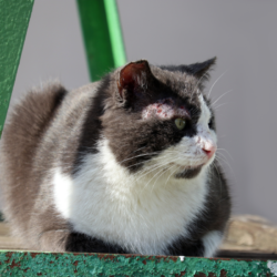
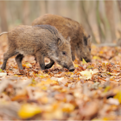
Canids, particularly dogs, as well as many domestic (sheep, cattle, horses, goats) and wild herbivores, and pigs can be infected.
Echinococcus granulosus infection occurs worldwide, with particularly active outbreaks in the Mediterranean Basin. In France, it is mainly found in the south and in Corsica (dog-sheep cycle), and more rarely in other regions (dog-oxen, dog-equid cycles).
It is transmitted via the digestive tra ct:
- Canids: by eating the organs or viscera of animals infected with Echinococcus granulosus. Canids harbour the worm in their small intestine and excrete its microscopic eggs in their faeces. The eggs adhere strongly to plants and soil and are highly resistant to environmental conditions.
- Herbivores: by ingesting food or water contaminated with canine excrement containing parasite eggs. These eggs are transformed into larvae that form cysts, initially in the liver.
Hydatidosis is often asymptomatic in canids. Symptoms in dogs and cats are virtually non-existent, and are referred to as healthy carriage. In the event of heavy infestation, diarrhoea and weight loss may be observed.
Dogs become infected by ingesting the viscera of cattle, sheep or other herbivores carrying echinococcus larvae. The adult parasites multiply in the dog’s digestive tract and their eggs are evacuated by the thousands in faeces. The eggs adhere to plants and can infect other herbivores, completing the parasite cycle.
How does contamination occur?
Hydatidosis is mainly transmitted via the digestive tract:
- Manual: By putting hands contaminated with parasite eggs on dogs’ fur, soil or plants into the mouth.
- Food: By ingesting contaminated plants (vegetables, fruit, wild berries and lettuce, mushrooms), eaten raw or undercooked.
- Accidental inoculation: By contact with cysts present on the organs or viscera of infected herbivores.
In France, cases of hydatidosis are rare and are mainly concentrated in the south-east and Corsica.
People engaged in certain activities are more exposed to the risk of contamination:
- Contact with dogs: shepherds, breeders, vets.
- Handling herbivore organs or viscera: slaughterhouse, rendering plant and veterinary laboratory workers.
- Working outdoors: contact with soil and low plants.
Dog tapeworms are transmitted by ingesting eggs after touching the coat of a dog contaminated with infected sheep viscera, or by ingesting water or raw vegetables contaminated with dog faeces. Cats can also transmit this parasite. Fox tapeworms can be caught by eating wild berries (raspberries, bilberries, wild strawberries) contaminated with fox faeces.
Dog tapeworms are found in regions where dogs and sheep coexist, particularly in the Mediterranean, South America, Australia, North Africa and Asia.
What are the symptoms of a hydatid cyst in humans?
Hydatidosis is often asymptomatic for several years. The organs most often affected are the liver and lungs. Treatment is mainly surgical and is generally very effective. The disease is often detected at the stage of complications due to compression or rupture of the cyst, leaving the intermediate host asymptomatic.
Liver hydatids often cause abdominal pain, nausea and vomiting. Liver cysts may present with abdominal pain or a palpable mass. Jaundice may occur if the bile ducts are obstructed. Rupture of the cyst into the bile duct, the peritoneal cavity or the lungs may lead to fever,urticaria or an anaphylactic reaction.
Lung cysts can cause haemoptysis, cough and dyspnoea. Although the infection is often contracted in childhood, it can remain latent for years, unless the cysts develop in vital organs.
Lung cysts can rupture, causing coughing, chest pain and haemoptysis. Hydatid cysts located in the liver and lungs are the most common, but they can also appear in the bones, kidneys, spleen, muscles and central nervous system. The asymptomatic incubation period may last several years before the cysts are sufficiently developed to trigger clinical signs.
In the case of pulmonary involvement, the clinical signs are chronic cough, chest pain and shortness of breath. Other signs include anorexia, weight loss and asthenia.
Unusual locations for cysts include the pleura, peritoneum, kidney, and less commonly the subcutaneous soft tissues, thyroid, pancreas, ovaries, joints and eyes. Medical imaging, such as ultrasound, CT and MRI, is essential for diagnosis, preoperative assessment and patient follow-up.
Zoom in on the structure of hydatid cysts
The pulmonary veins transport the embryos, which pass through the pulmonary filter to the left heart and then the aorta, joining the main circulation to settle in various organs. Once attached to an organ (liver, lung, etc.), the embryo is destroyed by an inflammatory response or develops into a cyst.
In the case of cyst formation, the embryo loses its hooks and becomes a protoplasmic mass, developing slowly. At one year, it becomes fertile and contains protoscolexes. The hydatid larva is made up of several layers:
- Adventitia (pericyst): fibro-conjunctive shell surrounding the parasite, highly vascularised, which eventually calcifies.
- Cuticle: resistant, hyaline membrane, allowing osmotic exchanges.
- Proligeral membrane: internal membrane rich in nuclei, responsible for the growth and reproduction of the cyst.
- Hydatid fluid: clear, aseptic, very rich in protoscolex, with antigenic and toxic properties.
- Hydatid sand: parasitic elements bathed in hydatid fluid, including protoscolex, free hooks, proligeral vesicles and daughter vesicles.
In the lungs, the hydatid grows with the least resistance and remains spherical. It may become oval or pear-shaped depending on the obstacles encountered. Risks include:
- Bronchial compression with risk of atelectasis.
- Ischaemic necrosis and erosion of the bronchial wall, leading to fistulas, infections and bronchiectasis.
- Rupture: in the bronchus (72-87% of cases), pleura (2-10%), or rarely in a venous trunk or the pericardium.
In the liver, cyst growth (up to 15-20 cm in diameter) leads to disruption of adjacent structures. Complications include compression of the bile ducts and suprahepatic veins, and ruptures in the bile ducts, peritoneal cavity, vessels or hollow organs.
How is this disease diagnosed?
On ultrasound or CT scan, the hepatic hydatid cyst appears as a liquid cystic image, sometimes calcified, with flaky echoes (hydatid sand) or multiple daughter vesicles. Gharbi’s classification describes the different aspects according to their stage of development:
- Type I: simple, homogeneous cyst, trans-sonorous on ultrasound, hypodense on CT scan (0 to 20 HU). The wall is thick. On MRI, the cyst contents are hypo T1, hyper T2, and the wall is hypo T2.
- Type II: membrane detachment, visible mainly on ultrasound.
- Type III: “honeycomb” appearance with partitions and daughter vesicles.
- Type IV: heterogeneous cyst of higher density (pseudotumour).
- Type V: calcified block (death of the parasite). To be distinguished from the peripheral calcifications seen in types III and IV.
The main differential diagnosis is hepatic amebic abscess due to Entamoeba histolytica. The cystic fluid can be analysed by puncture and protoscoleces(echinococcus embryos) can be found. PCR analysis is also possible.
Liver hydatid cysts can be diagnosed using a serological test (Weinberg reaction), although this test is often falsely negative for pulmonary cysts.Eosinophilia is generally absent, except in cases of cyst rupture. CT, MRI and abdominal ultrasound can be pathognomonic for hepatic cystic echinococcosis in the presence of daughter cysts and hydatid sand, but simple cysts can be difficult to differentiate from benign cysts, abscesses or malignant tumours.
Serological tests (enzyme-linked immunosorbent assay, indirect haemagglutination) are sensitive in detecting infection. The presence of echinococcal antigens can be confirmed by immunodiffusion (arc 5) or immunoblot. The CBC can detect hypereosinophilia.
What treatment is used?
There are four types of treatment for cystic echinococcosis: surgery, antiparasitic drug therapy, puncture therapy and a wait-and-see attitude.
- Surgery: Surgical resection involves removing the cyst(s). This is sometimes a complex operation because of the organ involved and the size of the cyst. Surgery is the treatment of choice for complicated cysts (ruptured, with biliary fistulas, compressing vital structures, > 10 cm in diameter, superficial with a risk of rupture, or accompanied by extra-hepatic disease).
- Puncture treatment (PAIR): The PAIR technique (percutaneous aspiration, injection of scolicidal agents and reaspiration) is used to treat cysts under CT guidance. It is often combined with albendazole-based drug treatment (400 mg twice daily) to prevent metastatic infection. However, there is a risk of cyst rupture, which can lead to anaphylactic shock and/or secondary dissemination.
- Antiparasitic treatment: Drugs such as albendazole or praziquantel are used to treat small unilocular cysts (< 5 cm) and inoperable cysts. Albendazole is administered one week before, during and at least 4 weeks after surgery or PAIR, for up to 6 months depending on clinical response. The cure rate with albendazole alone is around 30%.
- Observation: Simple surveillance may be proposed for asymptomatic cysts that may become inactive naturally. This type of attitude is adopted when cysts cause no symptoms.
In extreme cases, such as echinococcosis caused by the fox tapeworm, a liver transplant may be necessary to save the patient. Treatment then involves surgical removal of the cysts and prescription of antiparasitic drugs.
How can contamination be prevented?
To prevent echinococcosis in animals:
- Do not feed dogs with cyst-bearing viscera.
- Destroy infected viscera by rendering or deep burial.
- Systematically deworm dogs with an effective dewormer, while collecting and disposing of their faeces by burial.
Preventive measures for humans include :
- Training and informing employees about the risks associated with hydatidosis, hygiene rules, and preventive measures.
- Implementation of appropriate measures:
- Drinking water, soap, single-use paper towels and a first-aid kit.
- Appropriate, clean and well-maintained work clothes and personal protective equipment.
Hygiene rules include :
- Washing hands with soap and drinking water systematically after contact with animals, animal waste or excrement, before meals, breaks and at the end of the working day.
- In the event of a wound: wash, soap, rinse, disinfect and cover with an impermeable dressing.
- Regularly clean work clothes, gloves and boots.
In high-risk areas, you should :
- Wear gloves when worming dogs, then wash your hands.
- Avoid eating low-growing raw wild plants or fruit. Wash and cook them.
- Do not feed dogs raw offal.
- Follow basic rules of hygiene, such as cooking food thoroughly and washing hands before eating.
- Deworm dogs regularly with products such as praziquantel.
These simple methods have almost eliminated hydatidosis in New Zealand. Vaccines based on recombinant DNA are being developed in Australia for sheep. In poor communities, it is crucial to properly manage carcasses and viscera after family slaughter. Boiling infected livers and lungs for 30 minutes is a simple and effective method of destroying Echinococcus granulosus larvae.
Some epidemiological data…
Cystic echinococcosis is present on every continent except Antarctica. In endemic regions, incidence rates in humans can exceed 50 per 100,000 person-years, and prevalence can reach 5% to 10% in certain regions of Argentina, Peru, East Africa, Central Asia and China. In farm animals, the prevalence observed in slaughterhouses in hyperendemic areas of South America varies from 20% to 95%. Prevalence is highest in rural areas where the animals slaughtered are older.
Cystic and alveolar echinococcosis represent a major burden of disease. Worldwide, more than a million people are living with these diseases. Many of them will experience severe clinical syndromes without adequate treatment. Even with treatment, quality of life is often diminished.
For cystic echinococcosis, the post-operative mortality rate is 2. 2% on average, with around 6. 5% of cases relapsing after surgery. In 2015, the WHO estimated that echinococcosis caused 19,300 deaths and around 871,000 disability-adjusted life years (DALYs ) per year worldwide. The annual costs associated with treatment and losses to the livestock sector are estimated at 3 billion dollars.
The main outbreaks worldwide include the Mediterranean basin, North Africa, South America, Australia, certain regions of East Africa, Central Asia and Northern China. In Tunisia, echinococcosis is a hyper-endemic public health problem. In men, the annual surgical incidence is around 12.6/100,000 inhabitants. The serological and/or echographic prevalence of asymptomatic carriage varies from 1.5% to 4%.
In animals, canine parasitism by E. granulosus varies from 9.8% to 68. 4% depending on the region. Ewes can reach a prevalence of 70% in certain slaughterhouses. The main sites of echinococcosis in humans are the liver (60% of cases) and the lungs (20-40% of cases in adults, 47-77% in children).
Responses from the health authorities
Cystic echinococcosis is not a contagious animal disease. It is not a notifiable human disease and is not covered by an occupational disease table. Echinococcus granulosus is classified in hazard group 3 (R. 231-61-1 of the French Labour Code).
Robust surveillance data are essential for determining the burden of disease and assessing the effectiveness of control programmes. However, as with other neglected diseases affecting underserved populations, data are insufficient and require increased attention.
Monitoring cystic echinococcosis in animals is difficult because the infection is asymptomatic in cattle and dogs. In addition, communities or local veterinary services do not recognise the need for surveillance.
Cystic echinococcosis is a preventable disease because the definitive and intermediate hosts are domestic species. A programme combining vaccination of lambs, deworming of dogs and culling of older sheep could eliminate cystic echinococcosis in humans in less than ten years.
In 1995, the WHO set up the Informal Working Group on Echinococcosis to strengthen the fight against this disease. This group works with strategic partners to promote the collection and mapping of epidemiological data. It also aims to improve early diagnosis and clinical management.
Pilot programmes to combat echinococcosis are underway in several countries. Morocco, for example, has undertaken a project to decentralise diagnostic and therapeutic techniques. Similarly, Mongolia has set up a surveillance system to understand the burden of disease.
In collaboration with veterinary and food safety authorities, the WHO is supporting the development of programmes to combat echinococcosis as part of a “One World, One Health” approach. These efforts include deworming dogs and vaccinating lambs to break the transmission cycle.