Zoonoses are a group of infectious diseases that can be transmitted between animals and humans. This complex phenomenon raises major challenges in terms of public health, epidemiological surveillance and health risk management. From avian influenza to rabies and brucellosis, these diseases can have serious consequences for human health, the economy and the environment.
What is a zoonosis?
Zoonoses are diseases that can be transmitted between vertebrate animals and humans. They may be caused by bacteria, viruses or parasites. Transmission may be direct or indirect, via food or vectors such as insects. The World Organisation for Animal Health states that 60% of infectious human diseases are zoonotic. This includes zooanthroponoses and anthropozoonoses, for transmission from humans to animals and vice versa. These transmissions do not include non-infectious diseases, those induced in laboratories, transmitted passively by animal products, or common diseases with no interspecies transmission.
The health importance of zoonoses is increasing, with around 75% of emerging human diseases being zoonotic, affecting in particular certain professions in direct contact with animals. The medical consequences vary widely, from benign to fatal, with a significant economic impact, especially for livestock farming and public health budgets.
Eco-epidemiological models are used for risk assessment and warning systems, where the transition to major epidemics may depend on multiple factors. Human contamination can occur in the workplace, accidentally, during leisure activities or in the home.
Zoonoses can be classified as orthozoonoses, cyclozoonoses, metazoonoses and saprozoonoses, depending on the cycle of their pathogen. The epidemiological consequences vary, with some zoonoses causing no further transmission by infected humans, while others can spread further.
What are the means of prevention?
The prevention of zoonoses aims to interrupt the transmission of diseases from animals to humans. The priority is to act on the reservoir of the pathogenic agent, then on the worker’s exposure, and finally on the worker himself. Prevention measures, specific to biological agents and their mode of transmission, include work organisation, collective and individual protection, and staff information and training.
To control the reservoir, you need to :
- check the health of the animals
- quarantine new arrivals,
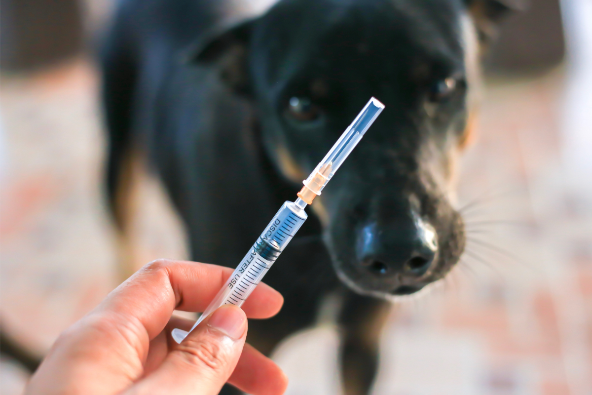
- vaccinate,
- treat sick animals,
- optimise rearing conditions,
- and prevent contact with wild animals, in particular by fencing.
Destroying the reservoir may require slaughtering animals or disinfecting them.
To reduce exposure, it is advisable to isolate sick animals, separate contaminated areas, limit access to high-risk zones, improve ventilation, reduce splashes and mechanise tasks. It is also important to clean and disinfect regularly, and to organise crawl spaces.
To protect workers, it is essential to provide them with suitable personal protective equipment. They must also be trained in their use and hygiene must be ensured with adequate facilities. Personal hygiene measures include washing hands, avoiding eye and nose contact with dirty hands, and disinfecting wounds.
Vaccination, while useful, is not a substitute for other preventive measures. Vaccination is subject to the worker’s agreement, following a risk assessment by the occupational physician.
Occupational health and safety departments play a key role in risk assessment, employee health monitoring and the management of incidents such as bites or scratches, with specific protocols for dealing with injuries.
Mycobacterium marinum skin infections
Mycobacterium marinum, an aquatic bacterium belonging to the atypical mycobacteria family, causes opportunistic infections in humans, including a rare disease known as aquarium granuloma, which affects people who come into contact with fish. This zoonosis, transmissible from fish to humans, is generally not fatal for immunocompetent individuals.
The bacterium, a bacillus measuring around 4 μm with a zebra-like appearance, lives in aquatic environments (fresh and salt water) and in various cold-blooded animals. Isolated for the first time in 1926, M. marinum was identified as a human pathogen in 1951. Infections, often linked to deteriorating aquatic conditions, affect more than 150 species of fish, with a variety of internal and external symptoms. In humans, it causes skin nodules, sometimes followed by ulceration, without fever or adenitis. Extracutaneous damage includes osteoarticular complications.
Diagnosis is difficult and delayed, based on the patient’s aquatic history and culture of the bacteria at 30°C. M. marinum is naturally resistant to antibiotics, but some, such as ethambutol and rifampicin, are preferred. Prevention is based on hygiene, aquarium maintenance and the use of gloves when handling the bacteria.
Aspergillosis
Brucellosis
Brucellosis, formerly known as Malta fever, is an anthropozoonosis caused by Brucella bacteria. Mainly transmitted to humans through contact with livestock or consumption of raw dairy products, the disease manifests itself as abortions in animals and recurrent fever, with joint or neurological complications in humans. Human-to-human transmission is very rare. Prevention efforts, including the control of livestock and the pasteurisation of dairy products, have significantly reduced its prevalence, especially in developed countries, where it is considered an occupational disease for certain exposed categories.
However, brucellosis remains a challenge in developing countries because of its socio-economic impact and its presence in various animal species. Treatment in humans is based on antibiotics, and management of the disease requires a tailored approach to avoid chronicity. Although the mortality rate is low, brucellosis is a major public health issue, requiring constant vigilance and prevention efforts, particularly in endemic regions.
Campylobacteriosis
Campylobacter, gram-negative bacteria that are widespread throughout the world, are pathogenic for livestock, causing disease and abortion in cattle, and transmissible to humans mainly through the ingestion of unpasteurised milk, contaminated water and undercooked meat. According to the EFSA, campylobacteriosis remains the main food-borne disease in the EU, mainly linked to chicken meat. Campylobacter jejuni and Campylobacter coli are the species most implicated in human cases, which can lead to autoimmune neuropathies such as Guillain-Barré syndrome.
Transmission is mainly dietary, with 20-30% of cases associated with the consumption of chicken meat. The infectious dose is low, and certain populations, such as children, the elderly and the immunocompromised, are more vulnerable. Symptoms include diarrhoea, abdominal pain and fever. Resistance to ß-lactam antibiotics requires treatment with macrolides or fluoroquinolones. Campylobacters are mainly commensals in the digestive tract of many animals, and contamination by humans or by animal contact is possible. In France, these bacteria cause around 493,000 infections a year, with around 15 deaths.
Anthrax
Anthrax is an acute infection caused by the bacterium Bacillus anthracis, affecting both animals and humans. Although it is rare in humans, it mainly affects herbivores. After the Second World War, experts considered Bacillus anthracis to be a potential biological weapon, and its notoriety increased following the attacks of 11 September 2001. Records of the disease date back to ancient times, when it was often confused with other animal diseases. In the 16th century, scientists officially recognised the disease, and in the 18th century identified its occupational transmission in humans. Major advances included the Rayer and Davaine rod distinction and Robert Koch ‘s work on spore formation.
As early as 1880, researchers developed veterinary vaccines, and then in the 20th century, they developed human vaccines, adapted to regional specificities. Bacillus anthracis is characterised by its resistance and ability to form spores, which contributes to its persistence in the environment. It has two major virulence factors: a capsule that prevents phagocytosis and two toxins that contribute to its virulence. It is mainly transmitted through contact with the spores of infected animals or their products.
Clinical forms in humans include cutaneous, gastrointestinal and respiratory manifestations, the latter being the most serious. The propagation of Bacillus anthracis as a biological weapon has been tried and tested, notably during the Second World War and in bioterrorist attacks in 2001. Treatment is based on the early administration of specific antibiotics, with recommendations issued by the CDC.
Choriomeningitis
Lymphocytic choriomeningitis is a viral disease caused by an arenavirus transmitted by rodents, identified mainly in the United States, with occasional cases in France. It often manifests as meningitis, characterised by fever, headache, nausea and sensitivity to light, and is generally of moderate severity. Infants may suffer more severe complications, such as microcephaly or hydrocephalus. Diagnosis is based on analysis of the cerebrospinal fluid, which is rich in lymphocytes, and is confirmed by RT-PCR or serology.
Vectors include house mice and other pet rodents, while dogs, cats and ruminants can be carriers without transmitting the virus. Human transmission comes mainly from contact with secretions or faeces from infected rodents, or by inhalation of contaminated aerosols. Symptoms in humans vary from asymptomatic forms to mild flu-like symptoms or more serious meningitis, with no permanent after-effects. The disease is rare, with few cases reported in France, and is not currently recognised as an occupational disease or a notifiable public health disease.
Cowpox
The cowpox virus, a member of the Poxviridae family and the Orthopoxvirus genus, mainly affects wild rodents, but also pets and cattle. It is distributed worldwide, with a poorly defined frequency in cattle in developed countries. Transmission occurs through direct contact between infected animals, mainly rodents and cats. Symptoms range from mild to fatal in rodents and include scabby lesions in cats.
In humans, transmission can occur through skin contact with an infected animal. The disease remains rare, with sporadic cases observed in Europe. Professionals in close contact with animals, such as pet shop staff, breeders and vets, are at increased risk. Symptoms in humans include a skin lesion that develops into a blackish crust, potentially accompanied by fever and muscle pain, with an increased risk of severe forms in immunocompromised or allergic individuals. Although it is not a contagious animal disease, it is a notifiable disease in humans, classified in hazard group 2 under the French Labour Code
Cryptococcosis
Cryptococcus neoformans is an environmental yeast present in two varieties: C. neoformans neoformans and C. neoformans gattii, causing cryptococcosis, an opportunistic disease in immunocompromised individuals and various mammals, particularly cats and ruminants. Transmission is mainly by inhalation of the yeasts, found in environments rich in organic matter such as bird droppings, especially pigeon droppings. The resistance of these yeasts in dry droppings is notable, lasting for several years. The disease manifests itself differently depending on the species: respiratory problems in cats, neurological problems in dogs and mastitis in ruminants, while pigeons remain asymptomatic.
In humans, cryptococcosis is seen almost exclusively in immunocompromised individuals, with around a hundred cases occurring each year in France. Symptoms mainly include damage to the central nervous system, and rarely to the skin or lungs. Human and animal contamination is global, with no inter-species transmission. Treatment varies according to severity, ranging from fluconazole alone to a combination of liposomal amphotericin B, flucytosine and fluconazole for cases of meningitis. Rising global temperatures could intensify the risk of infection, altering the epidemiology of this disease.
Cryptosporidiosis
Cryptosporidiosis, an intestinal disease caused by Cryptosporidium hominis and Cryptosporidium parvum, seriously affects animals (particularly cattle and birds) and is transmissible to humans. In young animals, it causes severe diarrhoea and intense weakness, often leading to death without effective treatment. In immunocompetent humans, it resolves in around ten days with symptomatic treatment, paromomycin being the reference molecule.
Parasites are inactivated by freezing or heating, but are resistant to most disinfectants, making chlorination of water ineffective. Transmission occurs mainly via contact with pets, excrement, or consumption of contaminated water or food. The disease is cosmopolitan, but the incidence varies, being higher in developing countries and among immunocompromised people.
Symptoms include profuse diarrhoea, abdominal pain, nausea and mild fever in humans, lasting three to fourteen days. There is no cure, but symptoms can be reduced with antibiotics. Prevention is based on food hygiene and protection of water resources.
Alveolar echinococcosis
Alveolar echinococcosis, also known as alveolar hydatidosis (AHD), is a disease caused by the parasite Echinococcus multilocularis. This parasite is mainly transmitted by foxes, affecting both certain carnivores (dogs, cats) and wild rodents, such as voles. The disease is endemic in the northern hemisphere, particularly in north-eastern France, the Massif Central and certain regions of Europe (Switzerland, Germany, Belgium, Italy).
It is transmitted to humans by ingesting plants contaminated with the parasite’s eggs, often without initial symptoms. If left untreated, alveolar echinococcosis results in high mortality due to progressive invasion of the liver. Early treatment with surgery and antiparasitics, in particular Albendazole for two years, generally leads to a cure. In the case of advanced disease, lifelong antiparasitic treatment is necessary to stabilise the condition. Thanks to these treatments, patients’ life expectancy can be almost normal.
Occupations at risk include contact with infected animals or contaminated land. A European surveillance network, based in Besançon, monitors the epidemiology of this rare but serious disease.
Contagious ecthyma or ORF
ORF is a zoonosis carried by the contagious ecthyma virus of the parapoxvirus family, mainly affecting sheep and goats, with possible cases in reindeer, camels and alpacas. The disease has a worldwide geographical distribution and is transmitted to humans mainly through direct contact with infected animals or contaminated material.
Symptoms in animals include crusty lesions on the skin and ulcerations of the mucous membranes, which can lead to death in young animals due to malnutrition. In humans, the infection manifests itself as an initial skin lesion (papule) that can develop into nodules, sometimes accompanied by fever in widespread cases. Farmers, vets and slaughterhouse staff are particularly at risk.
The disease is immunising, but recontamination is possible. The history of contact forms the basis of the diagnosis, which can be confirmed by biopsy. Treatment is limited to preventing superinfections. Vaccination of animals every 6-8 months is recommended.
Tick-borne encephalitis
Central European tick-borne encephalitis, belonging to the Flaviviridae family, affects both humans and animals. Infected species include wild and domestic mammals, birds and reptiles, the main vector being ticks of the genus Ixodes. Transmission is mainly by tick bite, with the possibility of transovarial transmission in ticks and, rarely, by consumption of infected raw dairy products. The geography of the infection is vast but poorly documented, extending across non-tropical Eurasia.
In animals, the infection is often asymptomatic, while in humans it manifests itself as “summer flu” symptoms and can progress to severe complications, such as neurological disorders or meningitis. The risk of exposure is higher for activities in wooded areas and for certain professions exposed to tick bites.
The severity of the disease varies according to the subtype of the TBEV virus (European, Siberian, Far Eastern), and the course can be serious, with cases of mortality and neurological sequelae. Although there is no specific treatment, the disease can be prevented by vaccination, which is recommended in high-risk areas.
Epidemiological understanding and risk management of tick-borne encephalitis require ongoing attention, particularly in view of the increase in the number of cases in Europe and changes in distribution areas due to climate change.
Bovine spongiform encephalopathy
Bovine spongiform encephalopathy (BSE), or mad cow disease, is a fatal degenerative disease of the central nervous system in cattle, caused by a prion protein. Discovered in Great Britain in 1986, it led to a major epizootic, mainly in the UK, between 1986 and the 2000s, with more than 190,000 cases.
The use of animal meal in cattle feed exacerbated the crisis, leading to interspecific transmission from scrapie in sheep or from an endemic origin in cattle. Consumption of contaminated cattle products also affected humans, with 231 victims showing symptoms similar to Creutzfeldt-Jakob disease. This crisis triggered an unprecedented ethical and economic awareness of farming practices. Despite the absence of treatment, the introduction of prophylactic measures has made it possible to contain the disease.
The pathogen, a prion protein, differs from viruses and bacteria in its resistance to conventional decontamination methods. Symptoms in cattle include cerebral changes and behavioural and locomotion disorders. Transmission to humans can lead to a form of Creutzfeldt-Jakob disease. The origins of the epidemic remain partially unexplained, with hypotheses ranging from interspecific contamination to mutations due to environmental factors. Cases of BSE following the ban on meat-and-bone meal (NAIF) suggest alternative transmission routes that have yet to be elucidated.
Mediterranean visceral leishmaniasis
Leishmania infantum, a protozoan parasite, is the causative agent of leishmaniasis in animals and humans, mainly around the Mediterranean basin, with some extension northwards. It is transmitted by the bite of sandflies. In animals, particularly dogs, and more rarely cats and certain wild carnivores, symptoms include a deterioration in general condition, weakness, thinness and skin lesions. Visceral leishmaniasis, or kala-azar, affects humans with symptoms such as irregular fever, emaciation and enlargement of the spleen and liver. In France, there are 20 to 30 cases a year, often linked to immunodepression.
The parasitic cycle sees the parasite migrate into the viscera, causing the death of the host without treatment. The species of Leishmania responsible vary according to geographical region. The spread of the disease in humans varies according to the immune response, with co-infection with HIV requiring particular attention. Diagnosis is based on microscopic visualization of amastigotes or serological tests, with PCR being the method of choice in immunocompromised patients.
Mediterranean spotted fever
Mediterranean spotted fever (MS F), a tick-borne vector-borne disease, is caused by the bacterium Rickettsia conorii and carried by the dog tick Rhipicephalus sanguineus. Prevalent on the French Mediterranean coast, it is most active in summer, although cases may emerge elsewhere following travel. Its eco-epidemiology remains unclear, but its geographical spread and severity appear to be increasing, classifying it as a re-emerging disease.
Identified for the first time in 1910 by Conor and Brüch in Tunis, MBF was known by various names before being unified under the term “Mediterranean spotted fever” in 1933. Its geographical distribution extends beyond the Mediterranean, also affecting Africa and Asia. It occurs seasonally, with a local incidence in endemic areas.
It is mainly transmitted by immature forms of the tick, which require prolonged contact to be infectious. The original reservoir may be wild rabbits, with other potential hosts identified.
Symptoms include a flu-like illness, a characteristic black spot at the bite site and, often, a skin rash. Although generally benign, severe complications can occur. Diagnosis is based on typical symptoms, confirmed by serology or PCR. Treatment is mainly antibiotic, aimed at reducing mortality, particularly in severe cases.
West Nile fever
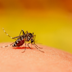
West Nile fever is caused by the West Nile virus, which is transmitted mainly by mosquitoes. 80% of infections are asymptomatic. When symptoms do appear, they include fever, headache, asthenia, nausea, vomiting, skin rash and lymphadenopathy. Serious cases can lead to meningitis or encephalitis, characterised by high fever, stiff neck, prostration, muscle weakness, tremors, convulsions, paralysis and, in some cases, coma. The risk increases with age and co-morbidities. Diagnosis is based on blood tests, and treatment is mainly symptomatic and may require hospitalisation. There is no vaccine for humans, but one is available for horses. Prevention is based on reducing the number of mosquito bites.
Birds act as a reservoir for the virus, which is transmitted to humans by mosquitoes that have fed on their blood. Human-to-human transmission is rare, but can occur via blood transfusions, organ transplants or from mother to child. Discovered in Uganda in 1937, the virus was detected in North America in 1999 and is also found in Europe, Africa, Asia and Australia.
Epidemiological surveillance is based on virus isolation from environmental samples, blood tests on wild birds, dogs and sentinel monkeys, and autopsies on dead birds. Mosquitoes, the main vectors, multiply under favourable conditions such as high temperatures or heavy rainfall. Individual prevention includes protection against bites and measures to control mosquitoes.
Q fever
Q fever, or coxiellosis, is a worldwide infectious disease caused by Coxiella burnetii. The main hosts are wild and domestic mammals, including cattle, sheep, goats, dogs and cats. It is transmitted mainly by inhalation of contaminated particles and contact with the secretions of infected animals. Its incubation period varies from 9 to 40 days, and a single bacterium can be sufficient for infection, making it a highly contagious zoonosis.
Historically, Q fever was identified in Brisbane, Australia, in 1935. Edward Holbrook Derrick initially called it “the Query fever”. The infectious agent was isolated by Derrick and Frank Macfarlane Burnet, the latter receiving the Nobel Prize in 1960 for his work. The study of this disease revealed wild mammals as natural reservoirs and ticks as transmission vectors.
Epidemiology shows that C. burnetii is present almost everywhere, except in Antarctica and New Zealand, with varied modes of transmission but rare human-to-human transmission. Contamination is more frequent in men, probably due to different occupational exposure and hormonal protection in women.
Pathogenesis involves C. burnetii entering host cells by phagocytosis, favoured by the acidic environment of the phagosome, allowing it to multiply. The bacterium can resist many environmental factors and antibiotics under certain conditions. Clinically, Q fever can manifest itself as a flu-like syndrome, atypical pneumonia, hepatitis and, in its chronic form, mainly as endocarditis. Effective treatment is based on cyclins, quinolones and hydroxychloroquine, with adaptations for pregnant women.
Animal scabies
Cat scratch disease
Cat scratch disease, also known as benign lymphoreticulosis or benign lymphogranuloma, is a bacterial zoonosis mainly transmitted by feline scratches. It is caused by bacteria of the Bartonella genus, mainly Bartonella henselae and, more rarely, Bartonella clarridgeiae. Cats, especially flea-infested strays, are the main vector. The disease is more common in hot, humid areas, often affecting children. It can be transmitted by scratching (75% of cases), biting (10%), or even without direct injury, via the cat’s saliva or contact with the eyes.
Symptoms begin with a papule at the site of inoculation, followed by lymphadenopathy, and may include myalgias, fever, headache, weight loss, skin rashes, and in rare severe cases, endocarditis or encephalitis. Diagnosis is based on clinical examination, serology, and sometimes PCR or lymph node biopsy. Treatment for severe forms includes antibiotics such as azithromycin. Precautionary measures such as avoiding scratching or biting, disinfecting wounds and flea control are recommended, given that there is no vaccine.
Avian flu or influenza
Avian influenza, or bird flu, is an infectious disease affecting birds, caused by A strains of the influenza virus. It varies from mild to fatal, and can lead to vast epidemics. The H5N1 strain in particular was identified as particularly dangerous to humans in 2004. It is mainly transmitted between poultry and, more rarely, to mammals, including humans, with a very low risk of contagion. Some birds carry the virus asymptomatically, and the virus has been detected in several mammalian species.
Historically, epidemics have affected bird populations since 1200 BC, with notable epizootics in Europe in the 17th and 18th centuries. Symptoms in birds include behavioural changes, respiratory problems and, in severe cases, neurological signs or sudden death.
Pathogenicity varies from strain to strain, with some being particularly lethal. Transmission to humans remains rare but has been documented, notably in fatal cases linked to H5N1 and H7N9. The fight against the disease includes epidemiological surveillance, precautionary measures for pet owners and official recommendations to prevent the spread of the disease. To date, no vaccine against avian influenza in humans is on the market, but vaccines are available for birds in epidemic areas.
Hantavirus
The genus Orthohantavirus, also known as hantavirus, belongs to the Hantaviridae family. The Hantaan virus is considered to be one of the most dangerous. These single-stranded RNA viruses with negative polarity belong to group V of the Baltimore classification. Humans, who are accidental hosts, can contract the virus from rodents, which vary from region to region: Apodemus spp. in Asia and the Balkans, Clethrionomys in Scandinavia and China, Peromyscus and Microtus in the United States, and Rattus spp. globally for the Seoul virus. Characterised by an envelope and a diameter of 180 to 115 nm, hantaviruses are responsible for haemorrhagic fevers and Hantavirus Pulmonary Syndrome (HPS), transmitted by inhalation of rodent excretions.
There are 25 antigenically distinct viral species. Human-to-human contagion is rare but has been documented. With no curative treatment available, prevention is based on reducing contact with rodents. Every year, around 200 cases of HPS occur, mainly in America, with a mortality rate of 40%, and 150,000 to 200,000 cases of haemorrhagic fever with renal syndrome worldwide, mainly in China. Diagnosis is based on the detection of antibodies, and treatment is symptomatic, as isolation is not necessary for European hantaviruses.
Monkey herpes B
The herpes B virus(Macacine alphaherpesvirus 1), belonging to the Simplexvirus genus and the Herpesviridae family, is a neurotropic pathogen for humans that can cause severe, often fatal, meningoencephalitis. Closely related to human herpes viruses types 1 and 2, its main reservoir is the macaque, where it is highly prevalent. Since its discovery, it has caused more than twenty human deaths. This underlines the importance of early diagnosis and treatment to increase the chances of survival. Otherwise, the mortality rate exceeds 70%.
Identified in 1932 after the death of Dr William Brebner, infected by a monkey bite, the virus was named Virus B by Dr Albert Sabin. It has specific virological characteristics. It has an enveloped linear double-stranded DNA and the ability to cross-react serologically with other herpesviruses. Its genome, fully sequenced in 2003, shows genetic similarities with HSV types 1 and 2. However, it differs in its ability to replicate in neurons.
Prevalent in Asian and African macaques, the herpes B virus is transmitted to humans mainly by bites, scratches or contact with infected secretions. Symptoms range from local itching to severe neurological complications if left untreated. Preventive measures include rigorous hygiene and antiviral treatment immediately after exposure. To date, no vaccine is available, and immunity against other forms of herpes offers no protection.
Hydatidosis
Hydatidosis, also known as hydatid echinococcosis or hydatid cyst, is an infection caused by ingestion of Echinococcus granulosus eggs, mainly through contact with dogs. This potentially fatal disease affects humans as well as many domestic and wild animals. The disease mainly develops in areas where dogs and herbivores live together.
The life cycle of the echinococcus requires definitive hosts (carnivores, especially dogs) and intermediate hosts (herbivores, and sometimes humans). Eggs ingested by the intermediate host release embryos that develop into cysts, mainly in the liver and lungs. If the intermediate host is eaten by the final host, the cycle continues.
Epidemiologically, echinococcosis affects 2 to 3 million people, with an annual cost of around 200 million dollars. Diagnosis is based on parasitological and serological methods, with techniques such as ELISA and Western Blot used to assess the presence of specific antibodies. Clinically, the disease is manifested by the formation of cysts, detectable by imaging (ultrasound, CT scan). Symptoms vary depending on the location of the cysts, often ranging from asymptomatic to rupture or compression of adjacent organs.
Leptospirosis
Leptospirosis is an infectious disease caused by the Leptospira bacteria, which is classified as an anthropozoonosis. They affect both humans and animals. Mainly transmitted through the urine of infected animals such as rodents, dogs and farm animals, these bacteria contaminate soil and water . They can lead to human infections without human-to-human transmission. The disease manifests itself through a wide variety of clinical signs, with a complex diagnosis due to the diversity of organs affected and the slowness of specific tests. Antibiotic treatment is nevertheless effective, and vaccination is recommended for certain occupational cases.
Historically, Adolf Weil described the severe form of leptospirosis in 1886, characterised by marked jaundice. In 1914, Inada and Ido discovered the bacterium L. icterohaemorragiae in Japan, and identified it as the initial causative agent. Over time, researchers discovered many similar bacteria, thus broadening the clinical and bacteriological spectrum of leptospirosis.
Human epidemiology shows a global presence of the disease, particularly in tropical areas. Risk factors vary from occupational to leisure activities involving exposure to contaminated water. The pathophysiology involves a generally cutaneous entry, followed by bacterial dissemination causing a variety of symptoms. Clinical forms vary from flu-like to severe, with multiple illnesses. Research is now focusing on understanding the variations in severity and developing more effective vaccines.
Listeriosis
Lyme disease
Lyme disease, also known as Lyme borreliosis, is a vector-borne zoonosis. It is transmitted to humans by the bite of Ixodes ticks. Researchers first identified Lyme disease in the American towns of Lyme and Old Lyme in 1975, mainly due to the bacterium Borrelia burgdorferi. In Europe, there is a greater diversity of borrelia. It includes Borrelia garinii and B. afzelii, causing a variety of clinical forms.
The disease initially manifests itself as migrant erythema around the bite. If left untreated, it can progress through three stages, affecting various systems and organs. There are acute or chronic cutaneous, articular or neurological forms. Antibiotics are effective in treating 90% of cases. On the other hand, the concept of “chronic Lyme disease” has given rise to debate about cases that have not been resolved by standard treatment.
Controversies over diagnosis and treatment are fuelling societal debates, particularly in the United States (Lyme War) and France (Scandale de Lyme). The evolutionary history of B. burgdorferi suggests that it has been present in North America for at least 60,000 years, with Ötzi, the first known human to be infected, having been infected around 5,300 years ago. The disease is expanding, becoming the most common vector-borne disease in the northern hemisphere. The complexity of co-infections and modes of transmission (mainly by ticks, but also potentially from mother to child) complicates the epidemiological landscape of the disease.
Ornithosis – Psittacosis
Ornithosis, also known as avian chlamydiosis, is an infection caused by the bacterium Chlamydophila psittaci of the Chlamydiaceae family. This disease, which includes psittacosis as a variant specific to Psittacidae, is a worldwide zoonosis that can be serious. Symptoms vary. They include fever, diarrhoea, conjunctivitis and respiratory disease. Their presence and severity depend on the infectious strain, the age and the species of the bird. The disease is transmitted by inhalation of dust contaminated with droppings or by the bite of infected birds, which may be asymptomatic.
These diseases are common in poultry farms, but rare in isolated birds. Cases of mammal-to-mammal transmission are extremely rare. The risks for humans include illnesses ranging from mild forms to severe atypical pneumonitis. The mortality rate falls to less than 5% under tetracycline antibiotic therapy. Ornithosis is a notifiable disease in several countries, in accordance with national and European regulations. It is recognised as an occupational disease in the poultry industry.
Pasteurellosis
Pasteurellosis is an infectious disease affecting animals and humans, caused mainly by Pasteurella multocida. This infection is frequently transmitted to humans by dog or cat bites or scratches, with a bacterial carriage rate of 40-50%. Symptoms appear rapidly, less than 24 hours after exposure, marked by intense pain and local inflammation. Treatment involves antibiotic therapy, typically cyclins or amoxicillin-clavulanic acid.
Various animal species, including game such as wild boar, can harbour these bacteria. In animals, pasteurellosis has a worldwide geographical distribution . Transmission is mainly via the respiratory tract or by bite, resulting in respiratory infections, abscesses and, in some cases, generalised infections.
In humans, in addition to transmission by bites and scratches, there is a risk through inhalation in confined spaces with infected animals. The frequency of pasteurellosis in humans is not well known . Some occupations at risk include veterinary surgeons, livestock farmers and abattoir workers. The disease manifests itself as painful oedema, fever and lymph nodes. It improves rapidly under antibiotic treatment, with rare complications involving joints or organs.
Rabies
Rabies is a viral encephalitis affecting mammals exclusively, and is almost always fatal after symptoms. This highly contagious disease is transmitted mainly by bite, affecting both animals and humans. The WHO estimates that there are around 59,000 deaths each year, mainly in Africa and Asia, with a prevalence among young people under the age of 15.
The symptoms, caused by a neurotropic virus, include neurological and behavioural disorders ranging from aggression to extreme calm. Vaccination of domestic animals and wildlife is essential to control this zoonosis. The rabies virus, which belongs to the Rhabdoviridae and Lyssaviruses, is sensitive to disinfectants and can mutate rapidly, easily crossing species barriers.
The reservoir of the virus appears to be certain bats, with transmission mainly via wild and domestic carnivores. Humans, considered accidental hosts, are rarely infected, with the majority of cases resulting from dog bites. Recovery is exceptional, except in certain bat species.
Prevention relies on vaccination and animal control measures to reduce transmission. Despite these efforts, global eradication remains a challenge, although significant progress has been made in certain regions, such as Europe, where rabies in foxes has been effectively controlled.
Red mullet
Rouget is a bacterial disease affecting mainly pigs, and occasionally lambs, calves and humans. This zoonosis is caused by Erysipelothrix rhusiopathiae. Historically, red mullet caused major damage in Europe and the United States in the 19th century. It led to the loss of millions of pigs. In 1881, under the direction of Louis Pasteur, Louis Thuillier isolated the bacterium responsible. This led to the development of a vaccine in 1883. Despite its current rarity in humans, the disease has been documented, with E. rhusiopathiae present in 30-50% of healthy pigs. Although rare, transmission to humans occurs mainly among professionals exposed to infected materials.
The disease presents three forms in pigs: acute, superacute and chronic, the latter being the least severe. In humans, diagnosis is based on cutaneous symptoms, and isolating the bacteria can be complex. The preferred treatment is benzathine benzylpenicillin or alternatives for those allergic to penicillin. Complications are rare, except in immunocompromised patients. Erysipelothrix rhusiopathiae is classified as a group 2 biological agent, with no reporting obligation for the disease.
Salmonellosis
Sodoku
Sodoku, a zoonosis transmitted by rat bites or scratches, can also be spread by ingesting contaminated water or milk. Rare in France, this disease is mainly seen in Japan, where it is caused by Spirillum minus. It is one of two forms of rat-bite fever, along with streptobacillosis, caused by Streptobacillus moniliformis. Historically, reports of the disease date back to ancient times in India. Notable cases were reported in the United States in 1839 and in Europe in 1884. Japanese research was prominent from 1890, identifying Spirillum morsus muris as the causative agent in 1916.
Before antibiotics, treatment was based on arsenic derivatives. Epidemiologically, rats are the main reservoir of the disease, which is often transmitted without any apparent symptoms in the animal. The disease manifests itself as inflammation at the bite site, followed by recurrent fever and skin rashes. Without treatment, the symptoms disappear and then reappear cyclically. Diagnosis is based on clinical observation and bacteriological tests, with PCR available. The treatment of choice includes penicillin or tetracyclines. Prevention requires rigorous hygiene and effective deratting.
Streptobacillosis
Rat-bite fever is a zoonosis caused by Streptobacillus moniliformis, transmissible to humans via rat bites or scratches. Transmission can be direct, through contact with the secretions of the infected animal, or indirect, via contaminated food and water. The risk of post-bite infection is 10%. Symptoms appear after an incubation period of 3 to 21 days, marked by fever, headache, chills, vomiting and acute arthritis, which may persist for several months. Petechiae may also appear.
In severe cases, the disease can lead to fatal endocarditis, pericarditis and tenosynovitis. Diagnosis is based on isolation of the germ or serological tests, although these are complex to carry out. Treatment with penicillin G, possibly supplemented by streptomycin, is effective. S. moniliformis infects various species and spreads globally, with no significant differences between strains. Reports of infectious episodes highlight the persistent health risk associated with this bacterium.
Streptococcus suis
Streptococcus suis infection, recognised as an occupational disease in France, mainly affects young piglets and, to a lesser extent, fattening pigs. Signs vary depending on the strain and the farm, with acute meningitis the most common manifestation. It is transmitted by skin penetration, aggravated by poor rearing conditions and stress. The management and prevention of asymptomatic carriers is complex. It requires particular attention to farming conditions and animal health. Rapid bacteriological diagnosis is crucial.
Given the high prevalence of S. suis in pigs and the difficulty of eradicating the bacterium, experts are targeting the major serotypes. Despite limited knowledge of pathogenesis, they have identified virulence factors, underlining the importance of monitoring antibiotic resistance.
Infection in humans occurs through contact with raw meat. Consumption leads to septicaemia, septic shock and meningitis, with significant cochleovestibular damage. Cases have been reported worldwide, with notable epidemics in China. In France, the risks mainly concern those working in the pig industry, although the exact route of transmission remains partially unknown. Illnesses such as pneumonia, endocarditis and arthritis can occur, requiring long courses of antibiotics. These can have serious after-effects.
Ringworm
Ringworm is a disease caused by dermatophyte fungi such as Microsporum or Trichophyton and their resistant spores. These diseases affect all mammalian species and, exceptionally, birds, with a worldwide distribution and a high frequency, especially in young people. Transmission occurs mainly through contact with infected animals or objects contaminated with spores, and rarely through soil. Symptoms vary depending on the fungus and the animal, generally including round, well-defined hairless areas.
In humans, transmission is similar, frequently affecting those in professional contact with animals. Symptoms include ring-shaped redness and inflammatory lesions, which heal after local and sometimes oral treatment. Historically documented, ringworm was common in Europe and remains endemic in some countries. Treatment has evolved, from X-rays and thallium salts to current recommendations including disinfection of personal items. The management of ringworm aims to control its spread and treat infections to reduce the risk of drug resistance.
Toxocariasis
Toxocariasis, or visceral larva migrans, is the most common helminthiasis in the world. It affects both developed and developing countries. This zoonosis is caused by ascarid larvae, mainly Toxocara canis (from dogs) and Toxocara cati (from cats). The larvae develop in tissues without reaching the adult stage outside their specific host. The parasite cycle involves the laying of eggs by the definitive host. These eggs evolve in the soil. The intermediate host then ingests them. The larvae then migrate through various organs.
In humans,infection occurs mainly through ingestion of eggs contaminating vegetables or through contact with infected soil, particularly in children practising geophagy or playing in contaminated sandboxes. The disease can be asymptomatic or manifest itself through a variety of symptoms. Symptoms can range from asthenia, to allergic reactions, to visual problems in the case of ocular toxocariasis. Diagnosis is based on serology. Treatment includes prevention of re-infection and the use of anti-helminthics such as albendazole. Prophylactic measures, both individual and collective, are essential to control its spread.
Toxoplasmosis
Toxoplasmosis is an infection caused by the protozoan Toxoplasma gondii . It mainly affects warm-blooded animals and humans, with certain felids, including cats, as the definitive host. Most cases in immunocompetent humans are asymptomatic. However, the infection can be serious in pregnant women, HIV-positive individuals and those with weakened immune systems. Overall, a third of the population is infected, with prevalence varying from region to region. Transmission can occur from mother to foetus, with risks varying according to the stage of pregnancy.
Discovered in 1908, T. gondii has a complex life cycle, alternating between asexual and sexual forms, mainly in cats. Humans can become infected through ingestion of infected raw meat, contact with cat faeces, or congenital transmission. Management includes prevention, especially in pregnant women, and appropriate treatment in the event of infection, particularly in immunocompromised patients and newborns with congenital toxoplasmosis. Professionals in contact with animals, raw meat or contaminated soil are particularly at risk.
Tuberculosis
In humans, transmission mainly results from inhaling contaminated aerosols, handling infected objects or ingesting raw milk. In mainland France, around fifty cases of tuberculosis of animal origin are recorded each year, among the 6,000 to 7,000 new cases of tuberculosis through human contamination. Occupations exposed to the risk of contamination include contact with live or dead animals, veterinary surgeons and abattoir workers. Most cases of M. bovis tuberculosis occur outside the lungs, particularly in the kidneys, with discrete initial symptoms.
Prevention of bovine tuberculosis is based on screening and eliminating sick animals. For a long time, the fight against human tuberculosis was based on BCG vaccination. However, given the limited effectiveness of this strategy, current prevention focuses on early detection and treatment of latent infection. Tuberculosis is a notifiable disease in France, Belgium and Switzerland. This enables rigorous epidemiological surveillance to be carried out.
Tularemia
Tularemia, a zoonosis caused by Francisella tularensis, affects around 300 species, including humans. Transmitted by direct contact with infected animals or by tick bites, it has a low incidence in France. A few dozen cases are recorded each year, mainly in the ulcero-ganglionic form. These cases also include serious pulmonary or septicaemic forms. Treatment is based on antibiotic therapy, adapted according to severity.
Historically, tularemia was potentially the first biological weapon in 1350 B.C. Officially described in 1911, the discovery of F. tularensis dates back to 1912, with subsequent studies establishing the role of ticks in its transmission. This highly infectious bacterium requires few germs to cause severe disease. It develops in macrophages and inhibits the host’s inflammatory response.
The symptoms vary: short incubation period, local forms with suppurated lymph nodes or typhoid, severe pneumonia, without specific signs. Diagnosis combines difficult culture, serodiagnosis and PCR, with treatment mainly using cyclins, fluoroquinolones and aminoglycosides. France considers tularemia to be a health hazard and a notifiable disease. It recommends epidemiological and health surveillance, including surveillance of wildlife. The bacterium represents a potential bioterrorism agent, classified by the CDC.


